- Table View
- List View

Life Cycle of a frog 1 of 5 (Frog ovum development) (UEB Contracted)
This page with five images illustrates stages in the development of a frog ovum. There is a locator dot shown, which will be at the top left of the page when the image is the right way up. The ovum is surrounded by a mass of jelly yolk, which is not shown. The top left of the page shows the zygote stage: a single cell with one nucleus at its centre, indicated by a dot. To the right of this is the cell in the process of division; it now has two nuclei. In the centre left of the page is the four cell stage with four separate cells, each with its own nuclei. Down the page from this the cells have divided again and now number sixteen. The bottom right section of the page shows the blastocyst stage. The cells have divided yet again and have formed a ball with a hollow containing fluid at its centre. There is a scale at the bottom of the page showing its actual size.
Life Cycle of a frog 1 of 5 (Frog ovum development) (UEB Uncontracted)
This page with five images illustrates stages in the development of a frog ovum. There is a locator dot shown, which will be at the top left of the page when the image is the right way up. The ovum is surrounded by a mass of jelly yolk, which is not shown. The top left of the page shows the zygote stage: a single cell with one nucleus at its centre, indicated by a dot. To the right of this is the cell in the process of division; it now has two nuclei. In the centre left of the page is the four cell stage with four separate cells, each with its own nuclei. Down the page from this the cells have divided again and now number sixteen. The bottom right section of the page shows the blastocyst stage. The cells have divided yet again and have formed a ball with a hollow containing fluid at its centre. There is a scale at the bottom of the page showing its actual size.
Life Cycle of a frog 2 of 5 (Frog spawn development) (Large Print)
On this page there are three images showing frog spawn development. There is a locator dot shown, which will be at the top left of the page when the image is the right way up. At the top of the page is the blastocyst surrounded by the jelly yolk in which it will develop. At the centre of the page is an image of the embryo stage; the cells are beginning to arrange themselves into the different areas that will become limbs and organs. The image at the bottom of the page shows a tadpole, still in the egg and surrounded by yolk, but ready to hatch. It has a scale to its right showing approximate size.
Life Cycle of a frog 2 of 5 (Frog spawn development) (UEB Contracted)
On this page there are three images showing frog spawn development. There is a locator dot shown, which will be at the top left of the page when the image is the right way up. At the top of the page is the blastocyst surrounded by the jelly yolk in which it will develop. At the centre of the page is an image of the embryo stage; the cells are beginning to arrange themselves into the different areas that will become limbs and organs. The image at the bottom of the page shows a tadpole, still in the egg and surrounded by yolk, but ready to hatch. It has a scale to its right showing approximate size.
Life Cycle of a frog 2 of 5 (Frog spawn development) (UEB Uncontracted)
On this page there are three images showing frog spawn development. There is a locator dot shown, which will be at the top left of the page when the image is the right way up. At the top of the page is the blastocyst surrounded by the jelly yolk in which it will develop. At the centre of the page is an image of the embryo stage; the cells are beginning to arrange themselves into the different areas that will become limbs and organs. The image at the bottom of the page shows a tadpole, still in the egg and surrounded by yolk, but ready to hatch. It has a scale to its right showing approximate size.
Life Cycle of a frog 3 of 5 (Tadpole development) (Large Print)
This is a multi-page image of the four stages of tadpole development, set on two pages. There are locator dots shown, which will be at the top left of each page when the images are the right way up. Each illustration has a scale showing its approximate size. Page 1: This page shows two illustrations of a tadpole with its head to the right of the page and its tail to the left. It is shown from the side so only one eye can be found. At the top of the page the tadpole is at an early stage of development. It still has gills to get its oxygen from the water, one of which can be found just to the left of its eye. At the bottom of the page the tadpole has grown and lost its gills. It has now developed so that it can breathe air through its mouth. Page 2: This page shows two more stages of development of the frog tadpole with its head to the right and tail to the left. At the top of the page the tadpole is viewed from the side with only one eye visible. One of its recently formed back legs can be found along the bottom edge of its body and the little bud of one of the emerging front legs can be found to the left of its mouth. At the bottom of the page the tadpole is seen from above. At the right of the image both of the tadpoles eyes are on view. To the left of this its front legs can be found and further left its back legs and tail. It is beginning to change from its 'fishy' shape to one that is more froglike.
Life Cycle of a frog 3 of 5 (Tadpole development) (UEB Contracted)
This is a multi-page image of the four stages of tadpole development, set on two pages. There are locator dots shown, which will be at the top left of each page when the images are the right way up. Each illustration has a scale showing its approximate size. Page 1: This page shows two illustrations of a tadpole with its head to the right of the page and its tail to the left. It is shown from the side so only one eye can be found. At the top of the page the tadpole is at an early stage of development. It still has gills to get its oxygen from the water, one of which can be found just to the left of its eye. At the bottom of the page the tadpole has grown and lost its gills. It has now developed so that it can breathe air through its mouth. Page 2: This page shows two more stages of development of the frog tadpole with its head to the right and tail to the left. At the top of the page the tadpole is viewed from the side with only one eye visible. One of its recently formed back legs can be found along the bottom edge of its body and the little bud of one of the emerging front legs can be found to the left of its mouth. At the bottom of the page the tadpole is seen from above. At the right of the image both of the tadpoles eyes are on view. To the left of this its front legs can be found and further left its back legs and tail. It is beginning to change from its 'fishy' shape to one that is more froglike.
Life Cycle of a frog 3 of 5 (Tadpole development) (UEB Uncontracted)
This is a multi-page image of the four stages of tadpole development, set on two pages. There are locator dots shown, which will be at the top left of each page when the images are the right way up. Each illustration has a scale showing its approximate size. Page 1: This page shows two illustrations of a tadpole with its head to the right of the page and its tail to the left. It is shown from the side so only one eye can be found. At the top of the page the tadpole is at an early stage of development. It still has gills to get its oxygen from the water, one of which can be found just to the left of its eye. At the bottom of the page the tadpole has grown and lost its gills. It has now developed so that it can breathe air through its mouth. Page 2: This page shows two more stages of development of the frog tadpole with its head to the right and tail to the left. At the top of the page the tadpole is viewed from the side with only one eye visible. One of its recently formed back legs can be found along the bottom edge of its body and the little bud of one of the emerging front legs can be found to the left of its mouth. At the bottom of the page the tadpole is seen from above. At the right of the image both of the tadpoles eyes are on view. To the left of this its front legs can be found and further left its back legs and tail. It is beginning to change from its 'fishy' shape to one that is more froglike.
Life Cycle of a frog 5 of 5 (Adult frog) (UEB Contracted)
This page is filled with the image of an adult frog stretched out to its full length. It is seen from above with its head at the top and back legs at the bottom. There is a locator dot shown, which will be at the top left of the page when the image is the right way up. To the left is a scale showing the approximate size of its body. In the top centre of the page is the frog's upper lip with two eyes slightly down from this. The frog's front legs, extending out to hand-like feet, can be found to either side. The frog's rounded body is in the centre of the page with two lines in the middle indicating the boney structure of its back. The lower half of the image shows the frog's two well-muscled rear legs extending down from its body and ending in three-toed feet at the bottom of the page.
Life Cycle of a frog 5 of 5 (Adult frog) (UEB Uncontracted)
This page is filled with the image of an adult frog stretched out to its full length. It is seen from above with its head at the top and back legs at the bottom. There is a locator dot shown, which will be at the top left of the page when the image is the right way up. To the left is a scale showing the approximate size of its body. In the top centre of the page is the frog's upper lip with two eyes slightly down from this. The frog's front legs, extending out to hand-like feet, can be found to either side. The frog's rounded body is in the centre of the page with two lines in the middle indicating the boney structure of its back. The lower half of the image shows the frog's two well-muscled rear legs extending down from its body and ending in three-toed feet at the bottom of the page.
Section through a molar tooth (Large Print)
This is an image of a molar tooth. There is a locator dot shown, which will be at the top left when the image is thenbsp;correct way up. The image is surrounded by an image border. The top of the tooth is at the top of the page and the root and jawbone at the bottom of the page. The components are labelled. The enamel, the grinding surface, is the upper layer. Down from this is the dentine layer which is slightly softer and surrounds the inner core which is the soft pulp containing the nerve and blood vessels. Going down to the bottom of the page are the two roots of the tooth which hold it firmly in place in the jawbone. The nerves and blood vessels come from the ends of the roots and go off to the left.
Section through a molar tooth (UEB Contracted)
This is an image of a molar tooth. There is a locator dot shown, which will be at the top left when the image is thenbsp;correct way up. The image is surrounded by an image border. The top of the tooth is at the top of the page and the root and jawbone at the bottom of the page. The components are labelled. The enamel, the grinding surface, is the upper layer. Down from this is the dentine layer which is slightly softer and surrounds the inner core which is the soft pulp containing the nerve and blood vessels. Going down to the bottom of the page are the two roots of the tooth which hold it firmly in place in the jawbone. The nerves and blood vessels come from the ends of the roots and go off to the left.
Section through a molar tooth (UEB Uncontracted)
This is an image of a molar tooth. There is a locator dot shown, which will be at the top left when the image is thenbsp;correct way up. The image is surrounded by an image border. The top of the tooth is at the top of the page and the root and jawbone at the bottom of the page. The components are labelled. The enamel, the grinding surface, is the upper layer. Down from this is the dentine layer which is slightly softer and surrounds the inner core which is the soft pulp containing the nerve and blood vessels. Going down to the bottom of the page are the two roots of the tooth which hold it firmly in place in the jawbone. The nerves and blood vessels come from the ends of the roots and go off to the left.
Section through an incisor tooth (Large Print)
This is an image of an incisor tooth. There is a locator dot shown, which will be at the top left when the image is the right way up. The image is surrounded by an image border.The top of the tooth is at the top of the page and the root and jawbone at the bottom of the page. The components are labelled. The enamel, the cutting surface, is the upper layer. Down from this is the dentine layer which is slightly softer. The inner core is the soft pulp which contains the nerves and blood vessels. Going down to the bottom of the page is the root of the tooth which holds it firmly in place in the jawbone. The nerves and blood vessels come from the end of the root and join the main vessels in the jaw shown in cross section as a dot.
Section through an incisor tooth (UEB Contracted)
This is an image of an incisor tooth. There is a locator dot shown, which will be at the top left when the image is the right way up. The image is surrounded by an image border.The top of the tooth is at the top of the page and the root and jawbone at the bottom of the page. The components are labelled. The enamel, the cutting surface, is the upper layer. Down from this is the dentine layer which is slightly softer. The inner core is the soft pulp which contains the nerves and blood vessels. Going down to the bottom of the page is the root of the tooth which holds it firmly in place in the jawbone. The nerves and blood vessels come from the end of the root and join the main vessels in the jaw shown in cross section as a dot.
Section through an incisor tooth (UEB Uncontracted)
This is an image of an incisor tooth. There is a locator dot shown, which will be at the top left when the image is the right way up. The image is surrounded by an image border.The top of the tooth is at the top of the page and the root and jawbone at the bottom of the page. The components are labelled. The enamel, the cutting surface, is the upper layer. Down from this is the dentine layer which is slightly softer. The inner core is the soft pulp which contains the nerves and blood vessels. Going down to the bottom of the page is the root of the tooth which holds it firmly in place in the jawbone. The nerves and blood vessels come from the end of the root and join the main vessels in the jaw shown in cross section as a dot.

The Goalkeeper's Revenge and other stories
by Bill NaughtonOne of a series of top-quality fiction for schools, this is a collection of stories of a Lancashire childhood, of football in the streets, fishing, fighting and school, of growing up and looking for work, and of characters such as Spit Nolan the champion trolley-rider, and Sam Dalt the goalkeeper.
New Windmills: The Goalkeeper's Revenge and other stories (PDF)
by Bill NaughtonOne of a series of top-quality fiction for schools, this is a collection of stories of a Lancashire childhood, of football in the streets, fishing, fighting and school, of growing up and looking for work, and of characters such as Spit Nolan the champion trolley-rider, and Sam Dalt the goalkeeper. Readership age: 10+.
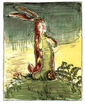
The Velveteen Rabbit (or How Toys Become Real)
by Margery Williams William NicholsonOriginally published in 1922, The Velveteen Rabbit has delighted young readers for nearly a century. The story follows a young boy who’s given a stuffed rabbit as a Christmas gift. After the rabbit befriends other nursery toys, he comes to the realization that he wants to become a real rabbit. Eventually, the boy becomes ill and is relocated; his room is then disinfected and all the boy’s toys are thrown out, including the velveteen rabbit. The rabbit sheds a real tear causing a fairy to appear and turn him into a real rabbit. This edition includes full-color illustrations, with image descriptions,from the original illustrator, William Nicholson. Each image accompanies the text to enhance young readers’ experience and immerse them in this captivating story. Reprinted hundreds of times since its initial publication, The Velveteen Rabbit is a timeless children’s classic lets young readers experience the true magic of friendship, love, and being honest with oneself. In 2007, the book was named one of "Teachers’ Top 100 Books for Children” by the National Education Association.
KS2 English SAT Buster Reading Answers (for Books 1-3) (PDF)
by Cgp BooksKS2 English SAT Buster - Reading Answer Booknbsp;