- Table View
- List View
Atlas of Fine Needle Aspiration Cytology
by Henryk A. DomanskiThis book covers all of the diagnostic areas where FNAC is used today. This includes palpable lesions and lesions sampled using various radiological methods, and correlations with ancillary examinations detailed on an entity-by-entity basis. As well as being a complete atlas of the facts and findings important to FNAC, this atlas is a guide to diagnostic methods that optimize health care. The interaction of the cytologist or cytopathologist with other specialists (radiologists, oncologists and surgeons) involved in the diagnosis and treatment of patients with suspicious mass lesions is emphasized and illustrated throughout. With contributions from experts in the field internationally and abundant colour images Atlas of Fine Needle Aspiration Cytology provides a comprehensive and up-to-date guide to FNAC for pathologists, cytopathologists, radiologists, oncologists, surgeons and others involved in the diagnosis and treatment of patients with suspicious mass lesions.
Atlas of Finger Reconstruction: Techniques and Cases
by Jian Lin Jianli Wang Deqing Hu Yongqing Xu Tianhao ZhangThis book covers latest advancement of finger reconstruction caused by severe injury leading to finger defects, presenting amount of valuable clinical experience and research achievement. This book provides practical guidance for hand and foot surgeons, micro- and reconstructive surgeons, trauma surgeons, orthopedic surgeons, general practitioners as well as trainees.The book mainly contains two parts: Part 1 focuses on development of the history of finger reconstruction, applied anatomy of the extremities, commonly used equipment and materials, frequently used medicines, preoperative treatment, selection of anesthesia, fundamental skill for finger reconstruction, postoperative treatment and management and methods of functional recovery after finger reconstruction. It is not uncommon in clinical scenarios that severe traumas caused by high energy damage in hands leading to life-long disability. The initial injury management is crucial. Surgeons face the arduous task of attempting to repair and reconstruct the injuries, reduce the patient’s disability and improve their quality of life. Part 2 demonstrates the concepts of different types of finger reconstruction with key points described by case presentations. Depending on the types and the severity of the lesions, accuracy of doctor’s judgment and proficiency of surgery skill have vital significance during the treatment of hand trauma together with patients’ compliance on functional rehabilitation.

Atlas of Forensic and Criminal Psychology
by Bernat-N. TiffonOriginally published in Spanish in 2017 by Libreria Bosch, Barcelona, the Atlas of Forensic and Criminal Psychology is a one-of-kind book made available in English for the first time. This unique work is highly illustrated with full-color images, providing a medico-legal examination of forensic pathology as it relates to cases of forensic psychological interest. The book begins with a historical perspective and includes images of patients to familiarize the reader with symptoms, the hazard-risk criteria, lethality, and suicidal rescue—research that Dr. Tiffon has addressed in his previous publications. Chapters present photographic records of cases to deepen forensic, psychologist, and medico-legal professionals’ insight into thoughts, behaviors, and mechanisms of self- and hetero-aggressiveness. Such cases illustrate the outcomes of various disorders manifested in individuals and victims; as such, they provide an understanding of the psychological-legal conclusions reached in such cases in order to adapt the legal and preventative measures for specific situations. Coverage includes affective, schizophrenic, and personality disorders as contributing elements in diagnostic judgments, noting the great difficulty such examples present to experts performing psychopathological evaluations after criminal, and often violent, events have occurred. Various psychopathological disorders are addressed as well as the technical treatment that should occur in each case from a psychological-forensic perspective. Features: • Presents a provocative look at various syndromes familiar to forensic psychologists, as applied to criminal cases and the pathology of suicide victims and homicide perpetrators • Combines the work of world-renowned expert contributors to examine the criminal, legal, and psychological facets of various diagnoses and case examples • Offers insight into the psychological state of suicide victims, considering their state of mind as a "psychological autopsy" In his previous books published in Spanish, Manual of Consulting in Psychology and Clinical, Legal, Legal, Criminal, and Forensic Psychopathology (2008), Manual of Professional Performance in Clinical, Criminal, and Forensic Psychopathology (2009), and the 4-volume Practical Criminological Atlas of Forensic Psychometry (2019-2020), Tiffon approached forensic psychology and psychopathology from a theoretical perspective. In the Atlas of Forensic and Criminal Psychology, his first book translated into English, Tiffon expands on these prior works, serving to provide a visual reference and guide to medical pathologists and consulting psychologists in cases of disorders in which psychopathological mutilation, injury, and self-injury occur.
Atlas of Forensic Pathology: For Police, Forensic Scientists, Attorneys, And Death Investigators (pdf)
by Joseph A. Prahlow and Roger W. Byard
Atlas of Frontal Sinus Surgery: A Comprehensive Surgical Guide
by David R. Lobo Jaime Viera Artiles Javier A. OspinaThe atlas offers a comprehensive and up-to-date overview of frontal sinus surgery. In recent years there have been great advances in endoscopic nasosinusal surgery but they have been particularly prominent in frontal sinus surgery. The book provides complete instructions for a gradual learning of the different surgical techniques and includes surgical pearls. It is enriched with videos presenting real-time guidance for frontal sinus endoscopic procedures. The book will meet the needs of both trainees and more experienced practitioners, and will enable them to make steady progress in endoscopic surgery and to adopt a more complete and safe approach to the frontal sinus. It will be of interest also for ophthalmologists, maxillofacial surgeons and neurosurgeons.
Atlas of Full Breast Ultrasonography
by Aristida Colan-GeorgesThis atlas describes and illustrates a novel approach, referred to as full breast ultrasonography (FBU), that represents a challenge to conventional breast imaging diagnosis. The coverage encompasses examination technique, diagnostic criteria, the imaging features of a wide variety of lesions, and role in follow-up. FBU involves anatomic ultrasound scanning based on the ductal echography technique proposed by Michel Teboul, supplemented by Doppler and real-time sonoelastography. The approach offers a variety of advantages. Compared with MRI it has a lower cost, wider availability, better resolution, and improved correlation with anatomy. Compared with mammography it has the benefits of absence of irradiation and pain, applicability in all cases, and better overall accuracy. Furthermore, the standardized technique of acquisition and interpretation means that it is suitable as a screening test, unlike classic ultrasonography. FBU is applicable in ultrasound BI-RADS assessment and is of value in depicting both benign and malignant conditions. It can be recommended as a first-line method of diagnosis and for the follow-up of treated breasts, regardless of the patient’s age, sex, or physical condition.
Atlas of Functional Anatomy for Regional Anesthesia and Pain Medicine: Human Structure, Ultrastructure and 3D Reconstruction Images
by Miguel Angel Reina José Antonio De Andrés Admir Hadzic Alberto Prats-Galino Xavier Sala-Blanch André A.J. van ZundertThis is the first atlas to depict in high-resolution images the fine structure of the spinal canal, the nervous plexuses, and the peripheral nerves in relation to clinical practice. The Atlas of Functional Anatomy for Regional Anesthesia and Pain Medicine contains more than 1500 images of unsurpassed quality, most of which have never been published, including scanning electron microscopy images of neuronal ultrastructures, macroscopic sectional anatomy, and three-dimensional images reconstructed from patient imaging studies. Each chapter begins with a short introduction on the covered subject but then allows the images to embody the rest of the work; detailed text accompanies figures to guide readers through anatomy, providing evidence-based, clinically relevant information. Beyond clinically relevant anatomy, the book features regional anesthesia equipment (needles, catheters, surgical gloves) and overview of some cutting edge research instruments (e.g. scanning electron microscopy and transmission electron microscopy). Of interest to regional anesthesiologists, interventional pain physicians, and surgeons, this compendium is meant to complement texts that do not have this type of graphic material in the subjects of regional anesthesia, interventional pain management, and surgical techniques of the spine or peripheral nerves.
Atlas of Functional Neuroanatomy
by Walter J. HendelmanUnderstanding how the brain is organized and visualizing its pathways and connections can be conceptually challenging. The Atlas of Functional Neuroanatomy, Third Edition addresses this challenge by presenting a clear visual guide to the human central nervous system (CNS). This edition has been completely reorganized to facilitate learning the stru
Atlas of Functional Shoulder Anatomy
by Giovanni Di Giacomo Nicole Pouliart Alberto Costantini Andrea De VitaThe anatomy of the shoulder is based on complex joint biomechanics. The purpose of this Atlas is to focus the reader’s attention on a series of bone, ligament, muscle and tendon structures and ultrastructures within the shoulder on which only the most recent international literature has reported in specialized journals. This Atlas also presents extremely high-definition images of "targeted" sections obtained from cadavers preserved using state-of-art techniques. This unique Atlas, making use of images of major visual impact, offers a scientific message on a topical joint, using simple but dedicated descriptive language.
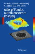
Atlas of Fundus Autofluorescence Imaging
by Frank G. Holz Steffen Schmitz-Valckenberg Richard F. Spaide Alan C. BirdThis lavishly illustrated unique atlas provides a comprehensive and up-to-date overview of FAF imaging in retinal diseases. It also compares FAF findings with other imaging techniques such as fundus photograph, fluorescein- and ICG angiography as well as optical coherence tomography. General ophthalmologists as well as retina specialists will find this a very useful guide which illustrates typical FAF characteristics of various retinal diseases.
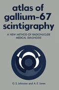
Atlas of Gallium-67 Scintigraphy: A New Method of Radionuclide Medical Diagnosis
by Gerald JohnstonIn 1970, under the sponsorship of Oak Ridge Associated Univer sities (ORAU), a group of clinical investigators formed the Cooper ative Group to Study Localization of Radiopharmaceuticals. The first radiopharmaceutical selected for study was 67-Gallium (67-Ga) administered as the citrate. The object of the study was to de termine the usefulness of 67-Ga in the diagnosis and treatment of patients with various malignancies. Funding for the project was granted by the U. S. Atomic Energy Commission and the National Cancer Institute, National Institutes of Health (NIH). The Nuclear Medicine Department of the Clinical Center, NIH, agreed to assist ORAU with aspects of this study, particularly with 67-Ga scin tigraphy of patients with lymphoma and Hodgkin's disease. Pre liminary reports from the ORAU study are in press. Since April 1971, 67-Ga scintigraphy has gained increasing use in the study of cancer patients at the Clinical Center, NIH, where well over 1000 such patients have been examined by this method. This monograph was written to present selected examples from this group of a variety of malignancies seen in this 28-month period. No attempt has been made to correlate this overall experience statistically. Rather, this presentation is to help familiarize the practitioner of Nuclear Medicine with the wide range of usefulness for 67-Ga scintigraphy while making him aware of the variation in scan appearance and watchful of the many pitfalls of 67-Ga scan interpretation. Permission to use these patient studies and x-rays was generously granted by Dr. Paul P.
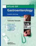
Atlas of Gastroenterology
by David H. Alpers Anthony N. Kalloo Neil Kaplowitz Chung Owyang Don W. PowellAccurate, high-quality images are especially vital for gastrointestinal therapy. The Atlas of Gastroenterology is a gold-standard tool that provides specialists with an outstanding array of images covering all facets of the field. With endoscopic ultrasonographs, computed tomography scans, magnetic resonance images, radionuclide images, and angiograms demonstrating every clinical condition from liver abscess, to endocrine neoplasms of the pancreas, to motility disorders of the esophagus, this atlas is simply a must-own resource for all gastroenterologists. Showing the range of the newest imaging technologies and incorporating over 1700 full-color images, this new edition is an ideal teaching tool, and the perfect companion to the Textbook of Gastroenterology.
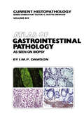
Atlas of Gastrointestinal Pathology: As Seen on Biopsy (Current Histopathology #6)
by M. Dawson T. MorsonBiopsy of the gastrointestinal tract has been revolution less busy) teaching hospital. These sort of techniques, which I confess interest me greatly because of the ized by the introduction of fibreoptics; the proximal additional information which they can yield when rightly reaches, as far as the second part of the duodenum, and chosen, are naturally linked with improved methods of the whole large bowel back to the caecum can now be tissue preservation in general, bearing in mind that the sampled under direct vision and multiple small biopsies need for special techniques often becomes apparent can be obtained. Only in the jejunum and ileum are there only when the biopsy has been conventionally still limitations on the sampling of localized as opposed to generalized conditions. The sheer volume of gastro processed and examined. However, I have firmly intestinal material passing through our own laboratories stabled this hobbyhorse and have included little that has risen steeply over the last years to form some 25% cannot be done in a district general hospital and nothing that I am not prepared to do myself. I have tried to of the total current work load and the rise continues; stress, particularly, common lesions which can cause nearly all of it is in biopsy form rather than as resected specimens.
Atlas of Gender and Health Inequalities in India (Demographic Transformation and Socio-Economic Development #16)
by Christophe Z. GuilmotoHow will the world's largest population approach its inequality challenges? This volume addresses this question by unraveling different strands of India's emerging health and gender geographies. It is the first book to offer a comparative study of these disparities in India, stressing the deep interaction between health challenges and patriarchal features. Most themes explored in this book illustrate the entangled nature of the social and regional determinants of gender and health imbalances in India. Through its rich cartography of contemporary India, the book represents the first Atlas exploiting district-level figures drawn from the latest sociodemographic survey conducted in 2019-21. After an initial methodological synopsis, the book is built around twenty chapters—illustrated by 75 original maps, figures, and tables prepared by thirty authors—and concludes with a synthesis of India's spatial patterns. Chapters engage with major themes of gender and health inequalities and explore an array of innovative indicators such as access to menstrual hygiene, cesarean deliveries, health insurance, son preference in fertility, female landownership, patrilocal systems, hypertension, anemia, hysterectomy, girl-only or single-child families, or traditional contraception. Together, they provide an often surprising glimpse into the present and future of India's gender and health landscape, highlighting the considerable progress accomplished over the last two decades alongside persistent gaps and emerging issues.
Atlas of General Surgical Techniques E-Book
by Courtney M. Townsend Jr. B. Mark EversAtlas of General Surgical Techniques covers the full spectrum and breadth of general surgery through nearly 1200 easy-to-follow anatomic drawings. Drs. Courtney M. Townsend, Jr. and B. Mark Evers present step-by-step guidance for common and complex procedures, including open and minimally invasive techniques. The highly consistent approach and format allow for large educational illustrations with pearls and pitfalls at the end of each chapter. Comprehensive coverage includes hot topics such as Thyroidectomy, Parathyroidectomy, Hepaticojejunostomy, Choledochojejunostomy, Splenectomy,Hernia Repair, Exploration of Neck for Trauma, and Subclavian Artery Stab. You’ll have a complete array of surgical procedures at your fingertips. 2009 PROSE Awards (awarded by Association of American Publishers for professional and scholarly excellence)Finalist/Honorable Mention, Clinical MedicineFeatures 1200 easy-to-follow, step-by-step anatomic drawings that clearly depict the full spectrum and breadth of surgical techniques—both open and minimally invasive. Covers hot topics such as Thyroidectomy, Parathyroidectomy, Hepaticojejunostomy, Choledochojejunostomy, Splenectomy, Hernia Repair, Exploration of Neck for Trauma, and Subclavian Artery Stab. Provides step-by-step instructions for each procedure in a highly consistent format that makes applying techniques easy. Highlights pearls and pitfalls at the end of each chapter so you know what to expect before entering the operating room. Presents the detailed guidance of authorities on what you need to know about common and challenging procedures.
Atlas of Genetic Diagnosis and Counseling
by Harold ChenThis book, Atlas of Genetic Diagnosis and understanding of these conditions and their care of Counseling, reflects my experience in 38 years of affected individuals and their families. It is also my clinical genetics practice. During this time, I have intention to bring the basic science and clinical m- cared for many patients and their families and taught icine together for the readers. Atlas of Genetic innumerable medical students, residents, and prac- Diagnosis and Counseling is designed for physicians ticing physicians. As an academic physician, I have involved in the evaluation and counseling of patients found that a picture is truly “worth a thousand with genetic diseases, malformations, and malfor- words,” especially in the field of dysmorphology. tion syndromes, including medical geneticists, Over the years, I have compiled photographs of my genetic counselors, pediatricians, neonatologists, patients, which are incorporated into this book to developmental pediatricians, perinatologists, obs- illustrate selected genetic disorders, malformations, tricians, neurologists, pathologists, and any phy- and malformation syndromes. A detailed outline of cians and health care professionals caring for each disorder is provided, describing the genetics, handicapped children such as craniofacial surgeons, basic defects, clinical features, diagnostic investiga- plastic surgeons, otolaryngologists, and orthopedics. tions, and genetic counseling, including recurrence I am grateful to many individuals for their risk, prenatal diagnosis, and management. Color invaluable help in reading and providing cases for photographs are used to illustrate the clinical fea- illustration.
Atlas of Genital Dermoscopy (Series in Dermatological Treatment)
by Giuseppe Micali Francesco LacarrubbaATLAS OF GENITAL DERMOSCOPY Edited by Giuseppe Micali, MD and Francesco Lacarrubba, MD Dermatology Clinic, University of Catania, Italy Dermoscopy, a non-invasive modern tool to enhance the diagnosis and monitoring of pigmented and non-pigmented skin disorders, is particularly suitable for use in the genital area, in which traditional invasive diagnostic procedures may be difficult or painful for the patient. Dermatologists, family physicians, and those involved in Sexual Health medicine will all benefit from this atlas showing the applications of dermoscopy in several external genital disorders both in males and females with large high-resolution color photographs throughout. Contents: Fordyce’s spots * Pearly penile papules and vestibular papillae * Genital warts * Molluscum contagiosum * Scabies* Pediculosis pubis * Candidiasis * Lichen planus * Lichen sclerosus * Lichen simplex chronicus * Zoon mucositis * Psoriasis * Vitiligo * Hidradenitis suppurativa * Melanosis * Dowling-Degos disease * Angiokeratoma * Lymphangioma circumscriptum * Melanocytic nevi * Seborrheic keratosis * Median raphe cyst * Squamous cell carcinoma in situ * Invasive squamous cell carcinoma * Extramammary Paget's disease * Melanoma
Atlas of Genitourinary Oncological Imaging (Atlas of Oncology Imaging #1)
by Ariadne M. Bach and Jingbo ZhangThe Atlas of Genitourinary Oncological Imaging presents a comprehensive visual review of appearances for normal anatomy and oncological diseases in the genitourinary system using over 900 radiological images and illustrations. The book presents current imaging techniques and discusses the role of imaging in pre-treatment staging and post-treatment follow-up. Diseases discussed include kidney, adrenal gland, upper tract, bladder, prostate, testes, and pediatric malignancies. Individual chapters include normal anatomy, imaging techniques, and pathology of each cancer type. The staging of the malignancy and what to include in the radiology report are discussed, and expected and complicated postoperative and post-treatment findings and recurrence are presented. Dedicated chapters on interventional and radiation therapy discuss their unique role in the management and treatment of oncology of the genitourinary system. Additionally, a chapter on chemotherapy toxicities discusses drug reaction treatment therapies unique to the genitourinary system.Edited and written by radiologists from the genitourinary disease management team at Memorial Sloan-Kettering Cancer Center, the Atlas of Genitourinary Oncological Imaging is an ideal resource for radiology and urology trainees seeking a review of the basics and for practicing radiologists looking for answers to challenging cases confronted in daily practice.
Atlas of Genitourinary Pathology
by Gregory T. MacLennan Liang ChengA single source of information about pathologic lesions of the adrenal, the urinary tract, and the male genital system, minimizing the need to consult numerous texts of limited scope, this book contains gross photos and photomicrographs of virtually every pathologic entity, and variants of those entities. The book is lavishly illustrated with images accompanied by text that explains the visual images, highlighting key diagnostic features and providing brief but helpful discussions of the differential diagnosis. This book is designed for practicing pathologists and pathologists in training as well as urologists, GU radiologists, GU radiation oncologists, and GU medical oncologists.
Atlas of Genodermatoses
by Gianluca Tadini Michela Brena Carlo Gelmetti Lidia PezzaniDiagnosing a genetic skin disease can sometimes be a difficult task for a dermatologist. This is especially true for genodermatoses-generally considered rare diseases seldom seen by practicing clinicians. As a result, professionals often have little experience with their diagnosis. The Atlas of Genodermatoses presents a unique collection of such ca
Atlas of Geriatric Dermatology
by Robert A. Norman Edward M. Young, JrThis is a comprehensive, practical, densely illustrated diagnostic and therapeutic guide for all geriatric dermatology providers. The book comprises 50 chapters and over 600 color photographs on topics ranging from common conditions such as basal cell carcinoma, rosacea, and seborrheic dermatitis to unusual conditions such as angiosarcoma, dermatofibrosarcoma protuberans, and porphyria cutanea tarda.Sections include:- Inflammatory conditions (including contact dermatitis, alopecia, erythema multiforme, pemphigus, bullous pemphigoid, porphyria, pruritus, psoriasis, rosacea, seborrhea, urticaria, xerosis, and more)- Infections (fungus, herpes simplex and zoster, scabies, lice, and warts)- Skin signs in systemic disease (skin tags, cutaneous metastases, xanthomas)- Regional dermatoses (intertrigo, leg ulcers, pressure sores)- Benign tumors (chondrodermatitis, cysts, ganglion, fibrous papule, seborrheic keratoses, lentigines, and benign vascular lesions)- Pre-malignant and malignant tumors (actinic keratoses, angiosarcoma, basal cell carcinoma, dermatofibroma and dermatofibrosarcoma protuberans, intraepidermal neoplasia, Kaposi's sarcoma, keratoacanthoma, lentigo maligna, cutaneous lymphoma, Mycosis fiungoides, melanoma, nevi and moles, and squamous cell carcinoma)