- Table View
- List View
Atlas of Endoanal and Endorectal Ultrasonography: Staging and Treatment Options for Anorectal Cancer
by Giulio A. Santoro Giuseppe Di FalcoWith contributions by numerous experts
Atlas of Endocrine Pathology (Atlas of Anatomic Pathology)
by Lori A. EricksonAtlas of Endocrine Pathology provides a comprehensive compendium of photomicrographs of common and uncommon entities in endocrine pathology. The volume includes histologic features of normal features, reactive conditions, hyperplasia and tumors. The most helpful diagnostic features are illustrated to provide direction and clues to the diagnosis of endocrine tumors. Furthermore, photomicrographs highlight the most pertinent diagnostic features in problematic diagnoses in endocrine pathology.Authored by a nationally and internationally recognized pathologist, Atlas of Endocrine Pathology is an important learning tool for those becoming familiar with the diverse entities encountered in endocrine pathology and a valuable reference for practicing pathologists faced with challenging diagnoses in endocrine pathology.
Atlas of Endometrial Histopathology
by Gisela Dallenbach-Hellweg Hemming PoulsenFor this second, expanded edition, most of the chapters have been extensively revised to include the latest immunohistochemical and molecular biological methods. With its clearly thought-out structure, this atlas enables the pathologist to find, classify, and differentially diagnose each disease. 280 top-quality illustrations - over half in full colour - are each accompanied by a description of the pathology of the disease, a summary of the causes, an explanation of its clinical importance, and treatment options, emphasising the importance of constant mutual co-operation with the attending gynaecologist. Microphotographs are arranged systematically according to morphology, and each is followed by a short description together with the aetiology and morphologic and differential diagnosis of the different conditions. Hemming Poulsen is head of the WHO Collaborating Center for the Histological Classification of tumours and the new chapter on neoplasms, for example, reflects this new WHO classification. Additional chapters cover gestational diseases as well as new findings in infertility and hormone replacement therapy.
Atlas of Endometrial Histopathology
by Gisela Dallenbach-Hellweg Dietmar Schmidt Friederike DallenbachThis new edition differs from the preceding ones in that there has been extensive revision of most chapters. Recent advances in research gained by immunohistochemical, molecular biological, and cytogenetic methods are included, as far as they are applicable for daily diagnostic work. The chapter on neoplasms, in particular, has been greatly expanded in accordance with the new WHO International Histological Classification of genital tract tumors, covering all pertinent differential diagnostic aspects. The chapter on malignant lymphomas and hematopoetic neoplasms involving the endometrium is a new contribution to this edition (F. D. , Ulm). We have revised the chapter on gestational diseases, incorpor- ing recent advances in the differential diagnosis of gestational trophoblastic tumors. New discoveries and experiences in correlating structure and function in infertility and in h- mone replacement therapy of peri- and postmenopausal patients are also included. Many new microphotographs have been added to illustrate the advances in tumor research and in immunohistochemical detection methods. We have updated the list of references including recent relevant publications. To the correspondents and consultants who have contributed valuable observations and suggestions, bringing thereby to our attention omissions in the second edition, we acknowledge our cordial thanks. We thank Dr. Wolfram Klapper, Director of the L- phom Register of the University of Kiel, for case material for making the micropho- graphic illustrations of lymphomas. The staff of Springer-Verlag has earned our gratitude for their skill in preparing this new edition.
Atlas of Endometriosis
by Caroline Overton Robert W. Shaw Lindsay McMillan Colin DavisEndometriosis affects women in the reproductive years, is associated with pelvic pain and infertility, and - although not life threatening - can seriously impair health, with huge economic and social consequences. It is arguably the most frequent problem encountered in contemporary More...gynecology and is the subject of much ongoing research and i
Atlas of Endomicroscopy
by Ralf Kiesslich Peter R. Galle Markus F. NeurathEndomicroscopy is a newly developed diagnostic tool that enables in vivo microscopy with subcellular resolution during ongoing endoscopy in the upper and lower gastrointestinal tract. It is a revolutionary technology. Endoscopy and pathology are no longer separate subjects and, as a result, endomicroscopy leads to a close interaction between endoscopist and pathologist. This Atlas of Endomicroscopy is the first book dealing with the new insights of endomicroscopy. It provides an overview about the development, the requirements, the technique, current indications and further possibilities of endomicroscopy.
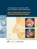
Atlas of Endoscopic Anatomy for Endonasal Intracranial Surgery
by Paolo Cappabianca Alessandra Alfieri Enrico de Divitiis Manfred TschabitscherIt is only recently that the use of the endoscope as the sole visualizing tool has been introduced in transsphenoidal pituitary surgery with its favorable related implications and minimal operative trauma. Of course, microscopic and endoscopic anatomy are basically the same, but the optical distorsion of endoscopic images is quite substantial compared to microscopic depictions. An endoscope lens produces images with maximal magnification at its center and severe contraction at its periphery. Nearer images are disproportionally enlarged and remote images are falsely miniaturized. This optical illusion may disorientate a surgeon who is not familiar with this peculiar condition at the skull base. This atlas acts as a guide through the endoscopic anatomy and gives detailed descriptions of the preoperative management and the surgical procedures.
Atlas of Endoscopic Major Pulmonary Resections
by Dominique GossotThe first version of this atlas was released as video-assisted major pulmonary resections were just emerging as a valid alternative to conventional techniques. In this second edition, many different techniques have been described, depending on the use or non-use of an accessory mini-thoracotomy and on the use or non-use of endoscopic instrumentation and video display. One of these techniques is the totally endoscopic approach, in which only endoscopic instruments and monitor control are used. This is the technique that will be described in this atlas. The purpose of this atlas is to describe each endoscopic pulmonary lobectomy and segmentectomy step by step, relying on brief technical notes and high-quality still pictures which are orientated and labeled to make them as comprehensible as possible. Each chapter is introduced by an anatomical background which is illustrated by three-dimensional reconstructions. Technical «tricks» and specific dangers are mentioned by pictograms. The technical descriptions of this atlas are based on the author’s technique, which can be different from other video-assisted approaches. Our intent is that surgeons embarking in video-assisted major pulmonary resections-whatever the approach they use-can find helpful hints and take their bearings in this totally new vision of pulmonary and mediastinal anatomy.Compared to the previous version, all chapters have been rewritten, taking into account the progresses of the technique and the technology and some new chapters have been added. Most steps of the procedures are now illustrated, not only with pictures, but also with a video clip.
Atlas of Endoscopic Major Pulmonary Resections
by Dominique GossotThis third edition Atlas of Endoscopic Major Pulmonary Resections describes in detail the totally thoracoscopic approach to major pulmonary resections, in which only endoscopic instruments and monitor control are used. Pulmonary lobectomies and segmentectomies are presented step by step, using brief technical notes and high-quality, clearly labeled still images. Each chapter begins with information on the anatomical background, which is illustrated in three-dimensional reconstructions. In turn, technical ‘tricks’ and specific pitfalls are explained in pictograms. The technical descriptions presented here are based on the author’s own technique, which in some cases differs from other video-assisted approaches. The goal is for surgeons embarking on video-assisted major pulmonary resections – regardless of which approach they use – to find helpful hints and guidance on this totally new vision of pulmonary and mediastinal anatomy.The first edition of this atlas was released at a time when video-assisted major pulmonary resections were just emerging as a valid alternative to conventional techniques. In the second edition, chapters on sublobar resections, as a new alternative to lobectomy in selected patients, were added. In this third edition, as the interest in sublobar resections is growing, and because they are challenging, the technique is dealt with in depth. In particular, readers will be introduced to new imaging technologies to support these techniques.
Atlas of endoscopic major pulmonary resections
by Dominique GossotIt is my greatest honor to be asked to write this foreword for the first edition of the Atlas of Endoscopic Major Pulmonary Resections by Dr Dominique Gossot. I have known Dr Gossot for over 15 years and have worked with him for many workshops and thoracic meetings. He is a pioneer in video-assisted thoracic surgery, and one of the most innovative thoracic surgeons I have known. Minimally invasive surgery has set a new standard of care for all surgical disciplines. Video-assisted thoracic surgery (VATS) offers a much kinder approach to the management of a wide variety of surgical conditions c- pared with conventional thoracotomy for these patients. Anatomical or major lung resections are a complex set of procedures commonly performed by thoracic s- geons. The adoption of the VATS approach for these procedures has received increasing acceptance by the thoracic surgical community, our pulmonologist and oncology colleagues, as well as the patients over the past two decades. There is now a growing body of evidence in the literature showing that the VATS approach is safe, oncologically sound, and associated with much lower morbidity compared with its conventional counterparts in the management of early lung cancers and benign conditions. Although there have been other books and atlases on VATS, this volume distinguishes itself in two respects.
Atlas of Endoscopic Neurosurgery of the Third Ventricle: Basic Principles for Ventricular Approaches and Essential Intraoperative Anatomy
by Roberto Alexandre DezenaThis book describes in practical terms the endoscopic neurosurgery of the third ventricle and surrounding structures, emphasizing aspects of intraoperative endoscopic anatomy and ventricular approaches for main diseases, complemented by CT / MRI images. It is divided in two parts: Part I describes the evolution of the description of the ventricular system and traditional ventricular anatomy, besides the endoscopic neurosurgery evolution and current concepts, with images and schematic drawings, while Part II presents a collection of intraoperative images of endoscopic procedures, focusing in anatomy and main pathologies, complemented by schemes of the surgical approaches and CT / MRI images.The Atlas of Endoscopic Neurosurgery of the Third Ventricle offers a revealing guide to the subject, addressing the needs of medical students, neuroscientists, neurologists and especially neurosurgeons.
Atlas of Endoscopic Perforator Vein Surgery
by John J. Bergan PeterGloviczkiA thorough description of new surgical treatment which accelerates the healing of formerly intractable venous ulcerations, and which can be carried out in a day-care surgical centre -- thus avoiding the need for hospitalisation. It treats the pathophysiology as well as the anatomy, and compares the results of surgical intervention to historical data. Lavishly illustrated by numerous colour photographs and line drawings.
Atlas of Endoscopic Plastic Surgery
by Edoardo RaposioConcentrating on technique, which is explained and illustrated in detail, this book is written by worldwide experts and provides detailed, step-by-step instructions on how to perform state-of-the-art endoscopic surgical techniques in the complex Plastic Surgery field. More than 300 high-quality photos help clarify complex techniques throughout the book. Atlas of Endoscopic Plastic Surgery represents a comprehensive description of the current endoscopic techniques in the plastic, reconstructive an aesthetic field. It supplies surgeons with all the information necessary to successfully accomplish an endoscopic approach to vary plastic surgery procedures, from carpal and cubital tunnel release, breast augmentation and reconstruction, migraine surgery, hyperhidrosis management, to facial aesthetic surgery, flap and fascia lata harvesting, and mastectomy and abdominal wall surgery.
Atlas of Endoscopic Sinus and Skull Base Surgery E-Book: Expert Consult - Online And Print
by Alexander G. Chiu James N. Palmer Nithin D AdappaGain a clear understanding of the entire spectrum of today’s rhinology and anterior skull base surgery with Atlas of Endoscopic Sinus and Skull Base Surgery, 2nd Edition. This thoroughly updated title increases your knowledge and skill regarding both basic or advanced procedures, taking you step by step through endoscopic approaches to chronic sinus disease, nasal polyps, pituitary tumors, cerebrospinal fluid leaks, sinonasal tumors, and more.Covers the full range of modern rhinology and anterior skull base surgery, from septoplasty and sphenoethmoidectomy to extended frontal sinus procedures, endoscopic craniofacial resections and complex skull base reconstructions.Clearly conveys the anatomy and detailed steps of each procedure with concise, step-by-step instructions; visual guidance features high-definition, intraoperative endoscopic photos paired with detailed, labeled anatomic illustrations.Includes new content on anterior skull base surgery that reflect new developments in the field.Helps you provide optimal patient care before, during, and after surgery with detailed information on relevant anatomy and surgical indications, instrumentation, potential pitfalls, and post-operative considerations.
Atlas of Endoscopic Sinus and Skull Base Surgery E-Book: Expert Consult - Online And Print
by James N. Palmer Alexander G. ChiuImprove your surgical outcomes with Atlas of Endoscopic Sinus and Skull Base Surgery by James N. Palmer, MD and Alexander G. Chiu, MD. Ideal for every otolaryngologist who performs basic or advanced rhinologic procedures, this beautifully illustrated atlas takes you step by step through endoscopic sinus and skull base surgeries as if the chapter authors were right there with you in the operating room.Consult this title on your favorite e-reader with intuitive search tools and adjustable font sizes. Elsevier eBooks provide instant portable access to your entire library, no matter what device you're using or where you're located.Benefit from the extensive knowledge and experience of leaders in the field as they walk you through endoscopic approaches to chronic sinus disease, nasal polyps, pituitary tumors, cerebrospinal fluid leaks, sinonasal tumors, and much more.Employ state-of-the-art techniques in your practice, from septoplasty and sphenoethmoidectomy to extended frontal sinus procedures, endoscopic craniofacial resections, balloon dilation, and complex skull base reconstructions.Visualize every step of each procedure thanks to high-definition, intraoperative endoscopic photos paired with detailed, labeled anatomic illustrations.Achieve optimal patient care before, during, and after surgery with detailed information on relevant anatomy and surgical indications, instrumentation, potential pitfalls, and post-operative considerations.
Atlas of Endoscopic Ultrasonography
by Frank G. Gress Thomas J. Savides Brenna C. Bounds John C. DeutschThe Atlas of Endoscopic Ultrasonography provides readers with a large collection of excellent images obtained from both diagnostic and therapeutic procedures. The Atlas includes a DVD which will be an invaluable addition to the library of trainee and practising gastroenterologists with video clips and searchable database of images. Together the book and DVD offer a first class collection of images to give a highly integrated introduction to endoscopic ultrasonography. The Atlas is an ideal companion to Dr Gress et al’s Endoscopic Ultrasonography, Second Edition.
Atlas of Endoscopic Ultrasonography
by Frank G. Gress Thomas J. Savides Brenna Casey Everson L. A. ArtifonAtlas of Endoscopic Ultrasonography Atlas of Endoscopic Ultrasonography Atlas of Endoscopic Ultrasonography, Second Edition offers an outstanding visual guide to this very common diagnostic and therapeutic endoscopic tool. With contributions from noted experts in the field, the Atlas contains 400 high-quality color and black and white images obtained from real cases, each accompanied by detailed annotation to aid readers in their understanding of this popular technical procedure. In addition, there is a companion website featuring 50 video clips of real-life procedures in action, as well as the entire collection of images from within the book. Updated throughout to include the most recent advances in interventional Endoscopic Ultrasound (EUS) guided therapies Contains a large collection of color images obtained from both diagnostic and therapeutic procedures, also available on the companion website image bank Provides a highly integrated and accessible multimedia introduction to endoscopic ultrasonography Includes a companion website offering insightful videos Written for gastroenterologists, students, residents, and radiologists, Atlas of Endoscopic Ultrasonography, Second Edition is an essential introduction to endoscopic ultrasonography.
Atlas of Endoscopic Ultrasonography
by Frank Gress Thomas Savides Brenna C. Bounds John C. DeutschThe Atlas of Endoscopic Ultrasonography provides readers with a large collection of excellent images obtained from both diagnostic and therapeutic procedures. The Atlas includes a DVD which will be an invaluable addition to the library of trainee and practising gastroenterologists with video clips and searchable database of images. Together the book and DVD offer a first class collection of images to give a highly integrated introduction to endoscopic ultrasonography. The Atlas is an ideal companion to Dr Gress et al’s Endoscopic Ultrasonography, Second Edition.
Atlas of Endoscopic Ultrasonography
by Frank Gress Thomas Savides Brenna Casey Everson L. A. ArtifonAtlas of Endoscopic Ultrasonography Atlas of Endoscopic Ultrasonography Atlas of Endoscopic Ultrasonography, Second Edition offers an outstanding visual guide to this very common diagnostic and therapeutic endoscopic tool. With contributions from noted experts in the field, the Atlas contains 400 high-quality color and black and white images obtained from real cases, each accompanied by detailed annotation to aid readers in their understanding of this popular technical procedure. In addition, there is a companion website featuring 50 video clips of real-life procedures in action, as well as the entire collection of images from within the book. Updated throughout to include the most recent advances in interventional Endoscopic Ultrasound (EUS) guided therapies Contains a large collection of color images obtained from both diagnostic and therapeutic procedures, also available on the companion website image bank Provides a highly integrated and accessible multimedia introduction to endoscopic ultrasonography Includes a companion website offering insightful videos Written for gastroenterologists, students, residents, and radiologists, Atlas of Endoscopic Ultrasonography, Second Edition is an essential introduction to endoscopic ultrasonography.
Atlas of Endoscopy with Narrow Band Imaging
by Manabu Muto Kenshi Yao Yasushi SanoWith its focus on narrow band imaging, this book is an excellent reference for new as well as experienced practitioners in the field of endoscopy. Narrow band imaging has brought about a revolutionary improvement in diagnostic endoscopy, enabling objective diagnosis and precise detection of lesions. It has enhanced the capability of endoscopy to facilitate qualitative diagnoses for the great benefit of patients who undergo endoscopic examinations. However, a standardized system of classification has not yet been established and many clinicians and researchers are not yet highly skilled in utilizing the technique or interpreting the images that are produced. This atlas addresses those issues, providing clear, simple and easy-to-understand descriptions illustrated with generous use of endoscopic images.
Atlas of Endovascular Venous Surgery E-Book: Expert Consult - Online
by Jose AlmeidaExpand your surgical skills with Atlas of Endovascular Venous Surgery, by Dr. Jose Almeida. This easy-to-access, highly visual reference offers the comprehensive, step-by-step guidance you need to achieve optimal outcomes in the surgical treatment of venous disorders. Detailed, full-color intraoperative illustrations and high-quality video clips capture key teaching moments, allowing you to better understand the nuances of surgery and equipping you to perform cutting-edge procedures and enhance your practice. The latest in laser-assisted distal stripping procedures, new devices for the treatment of venous reflux, and an evidence-based summary of the American Venous Forum guidelines are among many hot topics covered. What’s more, you can access the fully searchable, complete text - along with image and video libraries - online at www.expertconsult.com.See exactly how to proceed with high-quality, intraoperative videos that capture crucial teaching moments and provide a walkthrough of endovascular procedures. Access the full text, illustrations, video library, and more online at www.expertconsult.com. Get clinical anatomy reviews for each procedure through full-color illustrations depicting key anatomy and anatomic approaches to venous surgeries. Understand the correlation between imaging studies, anatomy, and surgical views by examining anatomy as seen in imaging modalities used during surgery - ultrasound, CT and MRI. Effectively apply the latest knowledge, techniques, and technologies including LADS (laser assisted distal stripping), new devices for the treatment of venous reflux such as ClosureFAST, evidence-based summaries of guidelines from the American Venous Forum, and more!
Atlas of Endovascular Venous Surgery E-Book
by Jose AlmeidaHighly visual and packed with useful, practical information, Atlas of Endovascular Venous Surgery, 2nd Edition, provides real-world instruction on the evaluation, diagnostic imaging, and medical and endovascular surgical management of acute and chronic venous diseases. Dr. Jose Almeida, pioneering expert in the field and host of the annual International Vein Congress, along with other highly regarded practitioners, offers an authoritative understanding of what causes increased venous pressure and solutions for reducing venous hypertension. Detailed, full-color intraoperative illustrations capture key teaching moments, helping you better understand the nuances of surgery and improve your ability to perform cutting-edge procedures.
Atlas of Enteroscopy: Endoscopy of the Small and Large Bowel; Retrograde Cholangio-Pancreatography
by L. Demling M. Classen P. FruehmorgenIt is my pleasure to introduce you to this new Atlas by Professor DEMLING and his colleagues on the very timely subjects colonoscopy, duodenoscopy and endoscopic cannu lation of the bile and pancreatic ducts. Professor DEMLING, unlike many others who carry his teaching and administra tive burdens, continues to be very personally involved in performing endoscopies. His clinic is one of the best organized and best equipped in the world. Professor DEM LING and his colleagues have been instrumental in introducing to the European continent these new techniques of duodeno scopy, colonoscopy and bile duct cannulation which were originally developed in Japan. They have added significant contributions of their own and now present to the reader a clear, concise, very well illustrated description of these methods. Dr. CLASSEN is one of the pioneers in endoscopic cannula tion. He has been kind enough to come to the United States and share his expertise with us at several Post Graduate Education programs. The students in these courses have been most enthusiastic about his presentations. Through this text he makes his extensive experience available to all endo scopists. The beginner at cannulation will find the illustrations of the various shapes the papilla may assume most helpful. I had the pleasure this summer of teaching a course in Brazil with Dr. FRUHMORGEN. Very few physicians have con centrated on colonoscopy to the extent that he has.
Atlas of Enteroscopy
by Gerard J. Gay, Francesco P. RossiniIn recent years, important technological innovations have made it possible to evaluate the small bowel endoscopically. Thus, traditional diagnostic methods such as imaging techniques are flanked by enteroscopy. The aim of this book is to present the results of enteroscopy as a method to diagnose small bowel disease, correlating it to the other investigative methods and above all to clinical findings. The book is a unique selected collection of images deriving from the results of documented experience and also including a brief clinical presentation, tables and diagrams of diagnostic algorithms. Renowned and accredited experts in gastroenterology, enteroscopy and researchers in the sector of small bowel disease have contributed to this volume.