- Table View
- List View
Atlas of Differential Diagnosis: MRI
by Guoguang FanAiming to equip readers with the knowledge required for accurate and timely diagnosis, this book presents illustrative cases that address a broad spectrum of clinical problems, including frequent, uncommon and rare diseases of brain, head and neck, spine, musculoskeletal system, chest and abdomen. To make readers understand easily, tips in differential diagnosis are summarized in the table, accompanying with typical images and key teaching points. It will be a valuable resource for diagnostic radiologists, residents, fellows, and related clinical doctors.
Atlas of Differential Diagnosis in Breast Pathology (Atlas of Anatomic Pathology)
by Puay Hoon Tan Aysegul A. SahinThis atlas illustrates the range of breast lesions with detailed correlation of gross and microscopic features. Where relevant, radiological images are incorporated. A description of normal, developmental and physiological breast morphology will serve as introduction to the main content of this atlas. Classification of tumors is based on the latest World Health Organization Classification of Tumors of the Breast, 4th edition, 2012. As immunohistochemistry is a key adjunctive tool in the workup of breast lesions as well as used in prognostic evaluation of breast cancers, appropriate examples are interspersed among the lesions where pertinent.
Atlas of Differential Diagnosis in Neoplastic Hematopathology
by Wojciech GorczycaManagement of tumor patients now relies on new individualized approaches to treatment, requiring extensive knowledge of the molecular makeup of tumors. Updated and expanded, the third edition of Atlas of Differential Diagnosis in Neoplastic Hematopathology examines not only the differential diagnosis but also the detailed morphologic, immunophenoty
Atlas of Differential Diagnosis in Neoplastic Hematopathology
by Wojciech GorczycaThis atlas presents not only the differential diagnosis but also the detailed morphologic, immunophenotypic, and especially genetic characteristics of the majority of hematolymphoid malignancies. An expert hematopathologist here provides a valuable resource to understand, use, or interpret one or more of these diagnostic modalities with confidence. This new edition has a compact format with up-to-date information - especially on genetic aspects - and will be an indispensable reference for all professionals in the specialty. *Provides an unrivalled visual resource for differential diagnosis in neoplastic hematopathology *Enables specialist and trainee oncologists and pathologists alike to understand, use, and interpret diagnostic modalities with confidence *Supplies quick access to information via tables, algorithms, and composite figures
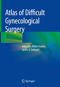
Atlas of Difficult Gynecological Surgery
by Anirudha Rohit Podder Jyothi G SeshadriCovering various difficult surgeries encountered in daily gynecological practice and providing unique insights into operative gynecology, this atlas offers an essential learning guide for gynecologists. Standard gynecological surgeries may turn out to be extremely complex on the operating table; this atlas demonstrates how the operating gynecologist can manage such procedures successfully without causing any injuries. For each surgery, it includes a series of color photographs accompanied by descriptive step by step notes, explaining to the readers the surgical steps and the problems that might occur if the dissection is not performed correctly. Helping readers understand and visualize the textbook descriptions of gynecological surgeries, the atlas offers a valuable resource for postgraduate students and fellows of obstetrics and gynecology, as well as senior practicing gynecologists, urologists and surgeons.
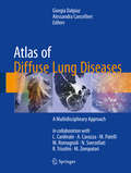
Atlas of Diffuse Lung Diseases: A Multidisciplinary Approach
by Giorgia Dalpiaz Alessandra CancellieriThis atlas is designed as an easy-to-use reference guide that identifies and illustrates the key patterns of diffuse lung diseases observed on high-resolution computed tomography (HRCT) and then documents in more detail the characteristics and appearances of the individual diseases, grouped on the basis of their prevalent pattern. A further feature of the book is its interdisciplinary nature: contributions from experts in various specialties are tightly interwoven throughout and many pathologic correlations are included. Less experienced readers will find that this atlas, with its wealth of figures and helpful color coding, steers them towards correct interpretation when confronted by the multiplicity and complexity of these diseases; those who are already experts, on the other hand, will benefit from the detailed coverage of individual diseases, which will deepen their understanding. At the end of the book, a graphically appealing and practice-oriented illustrated glossary with tips and tricks offers a further highly effective educational tool. Given the clinical spectrum of diseases covered, this atlas will prove invaluable for a wide range of healthcare workers, especially radiologists, pulmonologists, and pathologists.
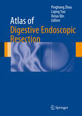
Atlas of Digestive Endoscopic Resection
by Pinghong Zhou Liqing Yao Xinyu QinDigestive Endoscopic Resection provides a minimally invasive method of diagnosing and treating gastrointestinal cancer. This book contains a comprehensive and detailed introduction to this technique. It includes preoperative preparation, surgical methods, intraoperative complications and postoperative management of advanced endoscopic procedures. It mainly focuses on endoscopic submucosal dissection (ESD) and endoscopic submucosal tunneling technologies, such as peroral endoscopic myotomy (POEM) and submucosal tunneling endoscopic resection (STER). This book presents essential surgical steps and diagnostic illustrations of these fast developing techniques. It provides a theoretical basis and valuable technical guidance for experts to perform ESD and summarizes this new treatment of gastrointestinal disease not only early cancers but also other gastrointestinal disorders including esophageal achalasia, etc. The Atlas of Digestive Endoscopic Resection will serve as an ideal reference for physicians and surgeons involved in endoscopic minimally invasive surgery.Editors Pinghong Zhou, Liqing Yao and Xinyu Qin are professors at Zhongshan Hospital affiliated to Fudan University, Shanghai, China.
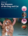
Atlas of Ear Diseases of the Dog and Cat
by Sue Paterson Karen TobiasBringing together a wealth of images of normal and diseased dog and cat ears, this is an indispensible diagnostic tool for the small animal veterinary practitioner seeing ear cases on a regular basis. This fully illustrated atlas covers the anatomy of the canine and feline ear, diagnostic techniques, a range of commonly seen diseases, and ear surgery. Atlas of Ear Diseases of the Dog and Cat is one of the most complete picture references for this rapidly expanding branch of small animal medicine and surgery. It is an invaluable aid for general practitioners, as well as those specialising in dermatology, and serves as an effective revision aid for veterinary students and those studying for further qualifications in veterinary dermatology. Includes over 400 high quality colour clinical images and clear line drawings Images are accompanied by clear explanatory text throughout Enables veterinarians to match cases seen in practice with photos supplied to aid diagnosis Written by highly qualified specialist veterinary dermatologist and veterinary surgeon
Atlas of Ear, Nose and Throat Pathology (Current Histopathology #16)
by L. MichaelsMany pathologists have little acquaintance with ear, nose concomitant biopsy have become commonplace in the and throat pathology. Some receive few specimens from management of throat disorders. It is hoped that. by ENT tissues; others are deterred from deeper study of the publication of this Atlas, pathologists receiving only material that emanates from regions the normal anatomy occasional specimens will be guided in their provision of of which is so forbidding in its complexity and holds no a report helpful to the clinician and those who are involved familiarity through autopsy investigation, for, apart from with a larger ENT service may be providec with a guide the larynx, there is usually no compelling indication for to the deeper understanding of the subject. examination of the ear, nose or throat at postmortem. Yet. The modern tendency in publication of ristopatholog equally with biopsy specimens from other parts of the ical microphotographs is to omit any statement of their body, the pathologist's report is consequential for the magnification, since it will usually be clear to the reader efficient handling of ear, nose and throat illnesses and what order of enlargement is involved. I n this Atlas, sometimes even for the patient's survival.

Atlas of Early Neoplasias of the Gastrointestinal Tract: Endoscopic Diagnosis and Therapeutic Decisions
by Frieder Berr Tsuneo Oyama Thierry Ponchon Naohisa YahagiThe latest edition of this text provides a comprehensive update on the current standards and newest skills in diagnostic endoscopy for pre/neoplastic lesions of the upper and lower gastrointestinal tract. The atlas outlines procedural requirements and strategies for detection and endoscopic staging (prediction of pT category) of small and minute early cancers, and presents endoscopic and high-resolution endosonographic criteria for submucosal invasiveness. The three major resection techniques, including risk profiles, and differential indications and contraindications for each technique are also outlined. In addition to thoroughly revised chapters from the previous edition, the atlas features new content on submuscosal neoplasias in the GI tract, new magnifying images of early gastric neoplasias, and new endoscopic images of adenoma, dyplasia, inflammatory bowel disease, and early cancer in the duodenum and small bowel.Written by experts in the field, Atlas of Early Neoplasias of the Gastrointestinal Tract: Endoscopic Diagnosis and Therapeutic Decisions, Second Edition is a valuable resource that will improve the diagnostic skills of endoscopists.

Atlas of Echocardiography in Pediatrics and Congenital Heart Diseases
by Maryam Moradian Azin AlizadehaslThis atlas provides a practical guide to the diagnosis of congenital heart disease using echocardiography in both adults and children. A plethora of high-quality echocardiography images provide practical examples of how to diagnose a range of conditions correctly, including aortic stenosis, tricuspid atresia, coronary artery fistula and hypoplastic left heart syndrome. Atlas of Echocardiography in Pediatrics and Congenital Heart Diseases describes the diagnostic management of a range of congenital heart diseases successfully in both adults and children. Therefore it provides a valuable resource for both practicing cardiologists who regularly treat these patients and for trainees looking to develop their diagnostic skills using echocardiography.

Atlas of EEG in Critical Care
by Lawrence Hirsch Richard BrennerAs the population ages, technology improves, intensive care medicine expands and neurocritical care advances, the use of EEG monitoring in the critically ill is becoming increasingly important. This atlas is a comprehensive yet accessible introduction to the uses of EEG monitoring in the critical care setting. It includes basic EEG patterns seen in encephalopathy, both specific and non-specific, nonconvulsive seizures, periodic EEG patterns, and controversial patterns on the ictal–interictal continuum. Confusing artefacts, including ones that mimic seizures, are shown and explained, and the new standardized nomenclature for these patterns is included. The Atlas of EEG in Critical Care explains the principles of technique and interpretation of recordings and discusses the techniques of data management, and 'trending' central to long-term monitoring. It demonstrates applications in multi-modal monitoring, correlating with new techniques such as microdialysis, and features superb illustrations of commonly observed neurologic events, including seizures, hemorrhagic stroke and ischaemia. This atlas is written for practitioners, fellows and residents in critical care medicine, neurology, epilepsy and clinical neurophysiology, and is essential reading for anyone getting involved in EEG monitoring in the intensive care unit.
Atlas of EEG in Critical Care
by Lawrence J. Hirsch Richard P. BrennerAs the population ages, technology improves, intensive care medicine expands and neurocritical care advances, the use of EEG monitoring in the critically ill is becoming increasingly important. This atlas is a comprehensive yet accessible introduction to the uses of EEG monitoring in the critical care setting. It includes basic EEG patterns seen in encephalopathy, both specific and non-specific, nonconvulsive seizures, periodic EEG patterns, and controversial patterns on the ictal–interictal continuum. Confusing artefacts, including ones that mimic seizures, are shown and explained, and the new standardized nomenclature for these patterns is included. The Atlas of EEG in Critical Care explains the principles of technique and interpretation of recordings and discusses the techniques of data management, and 'trending' central to long-term monitoring. It demonstrates applications in multi-modal monitoring, correlating with new techniques such as microdialysis, and features superb illustrations of commonly observed neurologic events, including seizures, hemorrhagic stroke and ischaemia. This atlas is written for practitioners, fellows and residents in critical care medicine, neurology, epilepsy and clinical neurophysiology, and is essential reading for anyone getting involved in EEG monitoring in the intensive care unit.

Atlas of EEG, Seizure Semiology, and Management
by Bassel Abou-Khalil Karl Edward Misulis Hasan Sonmezturk Kevin C. EssAtlas of EEG, Seizure Semiology, and Management is a comprehensive yet concise textbook with a focus on practical use of EEG and clinical neurology in the diagnosis and management of epilepsy. It begins with the physiological foundation of EEG, physics and electronics of EEG generation and recording, and technical details of EEG performance. Major sections then discuss seizure and epilepsy diagnoses, normal and abnormal EEG patterns correlated with clinical scenarios, guides to differential diagnosis of seizures, guides to medical and nonmedical management, and lastly teaching case studies. For this edition, there is a separate pediatric EEG and epilepsy section, in addition to discussion of pediatric aspects throughout the rest of the text. Revisions and additions have been made to keep up with the rapid pace of advancement in EEG and epilepsy. This new third edition is authored and edited by four senior faculty of Vanderbilt University Hospital and Vanderbilt Children's Hospital, all trained and board certified in EEG and Clinical Neurophysiology. These experts use their daily experience treating epilepsy in a busy academic medical center to provide a robust core of EEG and epilepsy knowledge to healthcare professionals who diagnose and manage epilepsy, and who are seeking to develop and enhance their knowledge of EEG performance and interpretation. New in this edition: · Compares the new epilepsy classification schemes with the old · Updated status epilepticus management, with new tables and flowcharts · New interpretations for a variety of EEG patterns, some of which have controversial implications

Atlas of EEG, Seizure Semiology, and Management
by Karl E. MisulisAtlas of EEG, Seizure Semiology, & Management, Second Edition, is a richly-illustrated guide to the performance and interpretation of EEG and management of epilepsy. Revised and updated in its Second Edition, this new text features hundreds of detailed EEGs, and covers the science in extensive scope and detail, beginning with basic electronics and physiology and then moving through EEG interpretation, epilepsy diagnosis, and ultimately epilepsy management. The new edition also includes all basic classifications and definitions of seizures and epilepsy, making it the perfect clinical companion. Atlas of EEG, Seizure Semiology, & Management utilizes full-color EEG presentations, alongside an easy-to-read synthesis of anatomy, physiology, and available treatment modalities. These detailed explanations of wave pattern, presentation, and treatment provide the student and practitioner with the most informed sense of clinical application and readiness. Atlas of EEG, Seizure Semiology, & Management covers every type of seizure, both epileptic and non-epileptic and divided into eight concise chapters. This unique atlas is necessary reading for all practicing neurologists, fellows, and residents.

Atlas of Elastosonography: Clinical Applications with Imaging Correlations
by Dirk-André Clevert Mirko D'Onofrio Emilio QuaiaWith the aid of an extensive series of high-quality images, this atlas describes all the potential applications of elastosonography in routine clinical practice. After a brief introduction on technical and physical aspects, the diagnostic benefits of elastosonography in a range of settings are illustrated with the aid of numerous representative cases. The coverage encompasses pathologies of the liver, spleen, pancreas, kidney, breast, thyroid, testis, musculoskeletal system, and vascular system. In addition, helpful correlations with imaging appearances on ultrasound and computed tomography are included. Elastosonography is a powerful, relatively new diagnostic technique that assesses the elasticity of tissues as an indicator of malignancy. Readers will find this book to be an excellent aid to use and interpretation of the technique.
Atlas of Elbow Surgery
by Andrea Celli Luigi CelliThis richly illustrated atlas is entirely devoted to the most modern surgical techniques of the elbow, offering a comprehensive, step-by-step and detailed examination of all the technical aspects of the surgical exposure of this joint. With the help of over 100 original drawings and photographs, it offers readers a unique understanding of elbow anatomy and surgical approaches, providing precise indications and surgical timing for clinical practice. An entire section focuses on the surgical management of the most common diseases of the elbow: the surgical exposures are related to pathologies affecting the lateral medial, anterior and posterior compartments of the elbow. This atlas will be of great value both to trainees and to specialists who manage disorders of the elbow, including orthopedic surgeons, traumatologists, and sports physicians, as well as anatomists.

Atlas of Electroencephalography in Sleep Medicine
by Hrayr P. Attarian Nidhi S UndeviaSleep Medicine is a field that attracts physicians from a variety of clinical backgrounds. As a result, the majority of sleep specialists who interpret sleep studies (PSG) do not have specialized training in neurophysiology and electroencephalography (EEG) interpretation. Given this and the fact that PSGs usually are run at a third of the speed of EEGs and that they usually have a limited array of electrodes, waveforms frequently appear different on the PSGs compared to the EEGs. This can lead to challenges interpreting certain unusual looking activity that may or may not be pathological. This Atlas of Electroencephalograpy in Sleep Medicine is extensively illustrated and provides an array of examples of normal waveforms commonly seen on PSG, in addition to normal variants, epileptiform and non-epileptiform abnormalities and common artifacts. This resource is divided into five main sections with a range of topics and chapters per section. The sections cover Normal Sleep Stages; Normal Variants; Epileptiform Abnormalities; Non-epileptiform Abnormalities; and Artifacts. Each example includes a brief description of each EEG together with its clinical significance, if any. Setting the book apart from others in the field is the following feature: Each EEG discussed consists of three views of the same page -- one at a full EEG montage with 30mm/sec paper speed, the same montage at 10mm/sec (PSG speed) and a third showing the same thing at 10 mm/sec, but with the abbreviated PSG montage. Unique and the first resource of its kind in sleep medicine, the Atlas of Electroencephalograpy in Sleep Medicine will greatly assist those physicians and sleep specialists who read PSGs to identify common and unusual waveforms on EEG as they may appear during a sleep study and serve as a reference for them in that capacity.

Atlas of Emergency Medicine Procedures
by Latha GantiThis full-color atlas is a step-by-step, visual guide to the most common procedures in emergency medicine. Procedures are described on a single page, or two-page spreads, so that the physician can quickly access and review the procedure at hand. The atlas contains more than 600 diagnostic algorithms, schematic diagrams and photographic illustrations to highlight the breadth and depth of emergency medicine. Topics are logically arranged by anatomic location or by type of procedure and all procedures are based on the most current and evidence-based practices known.