- Table View
- List View
Atlas of Diabetes
by Jay SkylerThis handbook is an invaluable resource for improving the management of diabetes. Chapters cover the fundamentals, including epidemiology, history and physical examination, and functional evaluations. Diabetes in children, adolescents, adults, and geriatrics are addressed. Differential diagnosis is emphasized, and evidence-based guidelines and patient-specific considerations aid the reader with injury evaluation and care. Notably, the book highlights the importance of understanding diabetic symptoms when determining the source of illnesses. In addition, the text presents the spectrum of treatment options for diabetes. The book is complete with appendices that explain the evidence-based approach used throughout and the science behind therapeutic modalities.
Atlas of Diabetes Mellitus
by Ian N. ScobieDiabetes mellitus is an extremely common disease that is reaching epidemic proportions owing to major increases in the incidence of obesity and the propensity for a sedentary life. The need for physicians to familiarize themselves with all forms of diabetes is essential and the Atlas of Diabetes Mellitus goes a long way toward making the task easie
Atlas of Diabetes Mellitus
by Ian N. Scobie David HopkinsThis completely revised and updated Fourth Edition of the Atlas of Diabetes Mellitus provides a broad coverage of all aspects of diabetes mellitus and an extensive collection of common and rare clinical images. It aims to provide an invaluable resource for anyone interested in the management of this ubiquitous clinical condition including primary care/ family physicians, endocrinologists, physicians in training, diabetic specialist nurses and other key professionals who are likely to be involved in the care of patients with diabetes mellitus.
Atlas of Diagnostic and Predictive Histopathology
by Shahid PervezThe second edition of this atlas covers the histopathology of all organ systems, supporting trainees and residents in the histopathological diagnosis of a wide range of inflammatory, neoplastic, degenerative, developmental and metabolic disorders. Though organ-specific books and atlases are increasingly available, there is a growing demand for a comprehensive guide to the histopathological diagnosis of each organ system, enabling histopathology trainees/residents and general pathologists to make reliable provisional or final diagnoses in routine practice. Each chapter covers a specific organ system and provides a better understanding of the microscopic pathology of common diseases and cancers, their interpretation, clinical presentation, epidemiology and demographics. The book also includes a chapter on ‘Predictive Pathology’ to help pathologists and other medical and healthcare professionals understand histopathology and report on it in keeping with standard international guidelines for individualized patient management. Intended as a concise and handy guide, this book offers a ready reference for trainees in histopathology, surgical pathology, anatomic pathology and cellular pathology, as well as practicing pathologists. It can also serve as a useful resource for biologists, biomedical scientists and physicians in general. The book features over 2200 illustrations with microscopic descriptions and interpretations. Besides H&E, special stains, IHC, FISH and radiology are included.
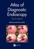
Atlas of Diagnostic Endoscopy, 3E
by Mohammad IbrarullahThis book is a compilation of endoscopic images of the upper gastrointestinal tract. The 3rd edition is enriched with high-resolution digital images highlighting the classification and staging of endoscopically relevant diseases. Serial documentation of diseases and procedures like corrosive injury, variceal obliteration, peptic ulcer etc. provides a complete, informative and interesting perspective. Rare conditions like Dieulafoy’s disease and Gastric antral vascular ectasia (GAVE) have been extensively discussed along with common diseases of the GI tract. This book outlines the technique and interpretation of endoscopic images proving to be a helpful guide to endoscopy practitioners. Key Features Explores various GI tract diseases through coloured, high resolution clinical photographs. Serves as a useful reckoner for trainee endoscopists and practitioners pursuing gastroenterology or gastrointestinal endoscopy. The text is updated with tables, flowcharts, classifications and international treatment guidelines.
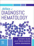
Atlas of Diagnostic Hematology E-Book
by Mohamed SalamaIdeal as a quick, easy-to-use reference in the laboratory or clinical setting, Atlas of Diagnostic Hematology is an abundantly illustrated guide to the vast range of malignant and non-malignant disorders of the blood. More than 1,200 vibrant, full-color images enable you to identify and compare the unique clinical and histologic features of various blood disorders and confidently reach a diagnosis. Coverage includes photos of actual cases that span the entire range of this complex field, including rare conditions and difficult diagnoses.Features more than 1,200 images including full-color pathologic and clinical images covering a wide range of hematologic malignant and non-malignant conditions. Covers a range of disease stages, from the slightest indication where diagnosis can be complicated or missed entirely, to what the average blood or biopsy sample signifying disease may look like, to an advanced stage where disease indications are abundantly clear. Helps you distinguish between similar and overlapping features and symptoms to arrive at an accurate diagnosis. Provides up-to-date information on infectious processes in blood and bone marrow, classification system of myeloid neoplasms, and indolent and aggressive mature T and NK-cell lymphomas. Includes diagnostic algorithms with differential diagnoses for conditions with similar histologic features and clinical symptoms. Contains the latest WHO classifications for pathologic, genetic, and clinical information.
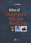
Atlas of Diagnostic Nuclear Medicine
by Anisah el HelouIntended for nuclear medicine specialists in training, it is equally an invaluable reference for other professionals and students. The richly illustrated chapters are devoted to individual organs and systems, with each chapter depicting the findings in selected pathological cases and in healthy individuals, with a comparison of nuclear medicine with other diagnostic imaging modalities. The full potential and also the limitations of modern nuclear medicine are described and sources of error are elucidated. The author is a well-versed nuclear medicine specialist with experience in research, teaching and clinical practice.

Atlas of Diagnostic Oncology E-Book
by Arthur T. SkarinAtlas of Diagnostic Oncology, 4th Edition, by Arthur T. Skarin, MD, FACP, FCCP, provides the guidance you need to diagnose a full range of neoplastic conditions with greater accuracy for better patient outcomes. An unrivaled collection of more than 2,500 images and drawings—combined with succinct, clinically focused text—equips you with essential information on pathology, diagnostic studies, staging, and clinical manifestations. New discussions on modern diagnostic PET imaging of cancer, and expanded coverage on the side effects of chemotherapy, bring you up to date on the issues impacting research and treatment. Expert Consult functionality—new to this edition—further enhances your reference power with convenient online access to the complete contents of the text, along with case studies that demonstrate effective approaches to diagnosis.A superb collection of more than 2,500 images encompasses the full spectrum of pathologic and radiologic studies, staging, and clinical manifestations, highlighting the pathologic anatomy of common clinical entities. A consistent chapter organization covers each disease’s incidence, epidemiology, etiology, and histopathology — as well as molecular biology, clinical features, diagnostic studies, and current clinical and pathologic staging — providing all the assistance you need to evaluate and monitor your patients effectively. This unique pictorial resource is a superb complement to treatment handbooks and major oncological texts. Expert Consult functionality provides online access to the complete text, fully searchable, with illustrations downloadable for your personal presentations, and case studies keyed to the book, at expertconsult.com. Completely updated chapters covering the newest genetic markers, imaging modalities, and pathologic techniques enable you to get the best results from today’s diagnostic tools. An expanded chapter on evaluating the side effects of chemotherapy, with additional images of reactions to the newest regimens, alerts you to potential common and uncommon side effects. Two new chapters address the complications of cancer and modern use of diagnostic PET scans, keeping you up to date on these hotly debated topics in the oncology community. A third new chapter covers malignant mesothelioma of the lung, plus other sites. Your purchase entitles you to access the web site until the next edition is published, or until the current edition is no longer offered for sale by Elsevier, whichever occurs first. Elsevier reserves the right to offer a suitable replacement product (such as a downloadable or CD-ROM-based electronic version) should access to the web site be discontinued.
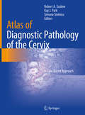
Atlas of Diagnostic Pathology of the Cervix: A Case-Based Approach
by Robert A. Soslow Kay J. Park Simona StolnicuThis book provides a practical guide to the diagnosis of cervical lesions. Chapters detail recent changes to diagnostic criteria and classification and the impacts these developments have on patient management. The anatomy and histology of the cervix are discussed, along with macroscopic and microscopic changes, prognostic and predictive parameters, epidemiological data, and staging systems. Atlas of Diagnostic Pathology of the Cervix: A Case-Based Approach utilizes diagnostic algorithms and highlights to offer readers appropriate management criteria and aims to give trainees, practicing pathologists, and gynecologists a case-based approach to the treatment of cervical lesions.
Atlas of Diagnostically Challenging Melanocytic Neoplasms
by Caterina Longo Giuseppe Argenziano Aimilios Lallas Elvira Moscarella Simonetta PianaThis atlas provides a clear, concise overview of the most challenging circumstances faced by clinicians and pathologists when dealing with melanocytic neoplasms. The book is structured as a case series; for each case, the clinical and dermoscopic appearances are presented, accompanied by a brief but comprehensive description and compelling histopathologic images. When available, in vivo confocal microscopy images are also included to highlight additional diagnostic clues. Identification of key messages and selected references will further guide the reader in the diagnosis and management of the neoplasm under consideration. It is well known that melanocytic lesions can be difficult to interpret. Some lesions show an ambiguous combination of morphologic criteria, and in these cases interpretation entails a high degree of subjectivity that results in low interobserver agreement even among expert pathologists. This atlas demonstrates how the addition of clinical information, including that provided by dermoscopy, can assist in reaching a more confident diagnosis.
Atlas of Differential Diagnosis: CT
by Guoguang FanAiming to equip readers with the knowledge required for accurate and timely diagnosis, this book presents illustrative cases that address a broad spectrum of clinical problems, including frequent, uncommon and rare diseases of brain, head and neck, spine, musculoskeletal system, chest and abdomen. To make readers understand easily, tips in differential diagnosis are summarized in the table, accompanying with typical images and key teaching points. It will be a valuable resource for diagnostic radiologists, residents, fellows, and related clinical doctors.
Atlas of Differential Diagnosis: MRI
by Guoguang FanAiming to equip readers with the knowledge required for accurate and timely diagnosis, this book presents illustrative cases that address a broad spectrum of clinical problems, including frequent, uncommon and rare diseases of brain, head and neck, spine, musculoskeletal system, chest and abdomen. To make readers understand easily, tips in differential diagnosis are summarized in the table, accompanying with typical images and key teaching points. It will be a valuable resource for diagnostic radiologists, residents, fellows, and related clinical doctors.
Atlas of Differential Diagnosis in Breast Pathology (Atlas of Anatomic Pathology)
by Puay Hoon Tan Aysegul A. SahinThis atlas illustrates the range of breast lesions with detailed correlation of gross and microscopic features. Where relevant, radiological images are incorporated. A description of normal, developmental and physiological breast morphology will serve as introduction to the main content of this atlas. Classification of tumors is based on the latest World Health Organization Classification of Tumors of the Breast, 4th edition, 2012. As immunohistochemistry is a key adjunctive tool in the workup of breast lesions as well as used in prognostic evaluation of breast cancers, appropriate examples are interspersed among the lesions where pertinent.
Atlas of Differential Diagnosis in Neoplastic Hematopathology
by Wojciech GorczycaManagement of tumor patients now relies on new individualized approaches to treatment, requiring extensive knowledge of the molecular makeup of tumors. Updated and expanded, the third edition of Atlas of Differential Diagnosis in Neoplastic Hematopathology examines not only the differential diagnosis but also the detailed morphologic, immunophenoty
Atlas of Differential Diagnosis in Neoplastic Hematopathology
by Wojciech GorczycaThis atlas presents not only the differential diagnosis but also the detailed morphologic, immunophenotypic, and especially genetic characteristics of the majority of hematolymphoid malignancies. An expert hematopathologist here provides a valuable resource to understand, use, or interpret one or more of these diagnostic modalities with confidence. This new edition has a compact format with up-to-date information - especially on genetic aspects - and will be an indispensable reference for all professionals in the specialty. *Provides an unrivalled visual resource for differential diagnosis in neoplastic hematopathology *Enables specialist and trainee oncologists and pathologists alike to understand, use, and interpret diagnostic modalities with confidence *Supplies quick access to information via tables, algorithms, and composite figures
Atlas of Difficult Gynecological Surgery
by Anirudha Rohit Podder Jyothi G SeshadriCovering various difficult surgeries encountered in daily gynecological practice and providing unique insights into operative gynecology, this atlas offers an essential learning guide for gynecologists. Standard gynecological surgeries may turn out to be extremely complex on the operating table; this atlas demonstrates how the operating gynecologist can manage such procedures successfully without causing any injuries. For each surgery, it includes a series of color photographs accompanied by descriptive step by step notes, explaining to the readers the surgical steps and the problems that might occur if the dissection is not performed correctly. Helping readers understand and visualize the textbook descriptions of gynecological surgeries, the atlas offers a valuable resource for postgraduate students and fellows of obstetrics and gynecology, as well as senior practicing gynecologists, urologists and surgeons.
Atlas of Diffuse Lung Diseases: A Multidisciplinary Approach
by Giorgia Dalpiaz Alessandra CancellieriThis atlas is designed as an easy-to-use reference guide that identifies and illustrates the key patterns of diffuse lung diseases observed on high-resolution computed tomography (HRCT) and then documents in more detail the characteristics and appearances of the individual diseases, grouped on the basis of their prevalent pattern. A further feature of the book is its interdisciplinary nature: contributions from experts in various specialties are tightly interwoven throughout and many pathologic correlations are included. Less experienced readers will find that this atlas, with its wealth of figures and helpful color coding, steers them towards correct interpretation when confronted by the multiplicity and complexity of these diseases; those who are already experts, on the other hand, will benefit from the detailed coverage of individual diseases, which will deepen their understanding. At the end of the book, a graphically appealing and practice-oriented illustrated glossary with tips and tricks offers a further highly effective educational tool. Given the clinical spectrum of diseases covered, this atlas will prove invaluable for a wide range of healthcare workers, especially radiologists, pulmonologists, and pathologists.
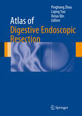
Atlas of Digestive Endoscopic Resection
by Pinghong Zhou Liqing Yao Xinyu QinDigestive Endoscopic Resection provides a minimally invasive method of diagnosing and treating gastrointestinal cancer. This book contains a comprehensive and detailed introduction to this technique. It includes preoperative preparation, surgical methods, intraoperative complications and postoperative management of advanced endoscopic procedures. It mainly focuses on endoscopic submucosal dissection (ESD) and endoscopic submucosal tunneling technologies, such as peroral endoscopic myotomy (POEM) and submucosal tunneling endoscopic resection (STER). This book presents essential surgical steps and diagnostic illustrations of these fast developing techniques. It provides a theoretical basis and valuable technical guidance for experts to perform ESD and summarizes this new treatment of gastrointestinal disease not only early cancers but also other gastrointestinal disorders including esophageal achalasia, etc. The Atlas of Digestive Endoscopic Resection will serve as an ideal reference for physicians and surgeons involved in endoscopic minimally invasive surgery.Editors Pinghong Zhou, Liqing Yao and Xinyu Qin are professors at Zhongshan Hospital affiliated to Fudan University, Shanghai, China.
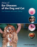
Atlas of Ear Diseases of the Dog and Cat
by Sue Paterson Karen TobiasBringing together a wealth of images of normal and diseased dog and cat ears, this is an indispensible diagnostic tool for the small animal veterinary practitioner seeing ear cases on a regular basis. This fully illustrated atlas covers the anatomy of the canine and feline ear, diagnostic techniques, a range of commonly seen diseases, and ear surgery. Atlas of Ear Diseases of the Dog and Cat is one of the most complete picture references for this rapidly expanding branch of small animal medicine and surgery. It is an invaluable aid for general practitioners, as well as those specialising in dermatology, and serves as an effective revision aid for veterinary students and those studying for further qualifications in veterinary dermatology. Includes over 400 high quality colour clinical images and clear line drawings Images are accompanied by clear explanatory text throughout Enables veterinarians to match cases seen in practice with photos supplied to aid diagnosis Written by highly qualified specialist veterinary dermatologist and veterinary surgeon
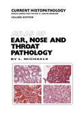
Atlas of Ear, Nose and Throat Pathology (Current Histopathology #16)
by L. MichaelsMany pathologists have little acquaintance with ear, nose concomitant biopsy have become commonplace in the and throat pathology. Some receive few specimens from management of throat disorders. It is hoped that. by ENT tissues; others are deterred from deeper study of the publication of this Atlas, pathologists receiving only material that emanates from regions the normal anatomy occasional specimens will be guided in their provision of of which is so forbidding in its complexity and holds no a report helpful to the clinician and those who are involved familiarity through autopsy investigation, for, apart from with a larger ENT service may be providec with a guide the larynx, there is usually no compelling indication for to the deeper understanding of the subject. examination of the ear, nose or throat at postmortem. Yet. The modern tendency in publication of ristopatholog equally with biopsy specimens from other parts of the ical microphotographs is to omit any statement of their body, the pathologist's report is consequential for the magnification, since it will usually be clear to the reader efficient handling of ear, nose and throat illnesses and what order of enlargement is involved. I n this Atlas, sometimes even for the patient's survival.