- Table View
- List View
Atlas of Cronkhite-Canada Syndrome
by Ryota Hokari Tadakazu HisamatsuThis manual treats both conceptual and practical aspects of gastrointestinal polyposis syndrome, which is characterized by dermatologic manifestations. The book offers both detailed and comprehensive analysis of the endoscopic findings achieved from 88 cases, which led to novel concepts and classification of the dermatologic manifestations according to distribution pattern, the thickness of mucosa, and intervening inflamed mucosa between polyps. The book also offers comparisons of the endoscopic features to pathological features. Abundant illustrations show the observations of the malignant lesions using special lights, such as NBI and BLI, to provide a comprehensive view of the Cronkhite-Canada syndrome (CCS) polyp.Atlas of Cronkhite-Canada Syndrome is essential guidance for gastroenterologists and pathologists who pursue the new diagnostic approach and treatment for CCS. It also attracts clinicians interested in the latest updates on this field and those who encountered the CCS patients with refractory to corticosteroid treatment. The risk of colorectal cancer in patients with CCS may increase because multiple inflammatory pseudopolyps make it more difficult to detect premalignant adenomas. Additionally, as endoscopy for GI disease becomes widespread in developing countries, the number of diagnosed patients will likely increase. Thus, it is crucial to have a broader understanding of the disease and the treatment strategy, and this book aims to simplify a complicated picture of the rare disease.
Atlas of Cryosurgery
by Nikolai N. KorpanThis atlas presents the fundamental aspects of modern cryosurgery. In the case of cancer patients it shows the discipline's advantages when compared to conventional surgical approaches. The text features definitions of the most frequently used terms, short descriptions of the historical and scientific background of cryosurgery, and an outline of cryosurgical equipment and techniques. Still further, the text covers the whole spectrum of experimental and clinical cryosurgery and the results of cryosurgical treatment of tumours in, for example, the liver, lung, and brain. Over 500 illustrations, mostly in colour, demonstrate the cryosurgical approach.
Atlas of CT Angiography: Normal and Pathologic Findings
by Gratian Dragoslav Miclaus Horia PlesThe second edition of this atlas presents a wealth of normal and pathologic findings observed on CT angiography with 3D reconstruction in diverse clinical applications, including the imaging of cerebral, carotid, thoracic, coronary, abdominal, and peripheral vessels. The superb illustrations display the excellent anatomic detail obtained with CT angiography and depict the precise location of affected structures and lesion severity. Careful comparisons between normal imaging features and pathologic appearances will assist the reader in image interpretation and treatment planning, and the described cases include some very rare pathologies. In addition, the technical principles of the modality are clearly explained and guidance provided on imaging protocols. This edition is the outcome of 18 years of work by a renowned radiological team whose research focuses specifically on vascular pathology of the whole body and the role of CT angiography in its assessment. Numerous new images are presented and three additional chapters address cerebral arteriovenous malformations, congenital cardiac malformations in children and CT venography. The atlas will be invaluable for radiologists, neurologists, neurosurgeons, cardiologists, cardiovascular surgeons, and medical students..
Atlas of CT Angiography: Normal and Pathologic Findings
by Gratian Dragoslav Miclaus Horia PlesThis atlas presents normal and pathologic findings observed on CT angiography with 3D reconstruction in a diverse range of clinical applications, including the imaging of cerebral, carotid, thoracic, coronary, abdominal and peripheral vessels. The superb illustrations display the excellent anatomic detail obtained with CT angiography and depict the precise location of affected structures and lesion severity. Careful comparisons between normal imaging features and pathologic appearances will assist the reader in image interpretation and treatment planning and the described cases include some very rare pathologies. In addition, the technical principles of the modality are clearly explained and guidance provided on imaging protocols. This atlas will be of value both to those in training and to more experienced practitioners within not only radiology but also cardiovascular surgery, neurosurgery, cardiology and neurology.
Atlas of Cutaneous Branch Territories for the Diagnosis of Neuropathic Pain
by Claude J. Spicher Tara L. Packham Nadège Buchet Isabelle Quintal Pierre SprumontThis atlas is the result of research involving over 3,000 patients consecutively recruited since 2004. Clinical practice gives the opportunity to observe many more Aβ axonal lesions (axonotmesis) than transections (neurotmesis), consequently the mapped hypo aesthetic territories are partial. Therefore, the authors define for each cutaneous nerve branch, the autonomous territory and the boundary markers of the largest territory of cutaneous origin. Each anatomical chart of a cutaneous branch is the superposition of tens, even hundreds of observations seen in clinical practice – based on 3,133 maps of observed cutaneous hypoaesthetic territories. The data collected has also been cross-referenced with that published in nearly 100 other anatomy books.This 1st English edition – based on the 3rd French edition published by Sauramps Medical – illustrates the usefulness of anatomical knowledge for clinical practice. More precisely, it seeks to demonstrate how these topographic elements can offer valuable support, both for the clinical anamnesis, and for the clinical examination of neuropathic pain patients. This atlas is at the crossroads between the medical and rehabilitation disciplines. Accordingly, it addresses the needs of medical doctors, from GPs to specialists, and of pain therapists, and offers a valuable asset for all health professionals who are dedicated to the management of pain and associated problems.
Atlas of Cutaneous Lymphomas: Classification and Differential Diagnosis
by Joi B. Carter Amrita Goyal Lyn McDivitt DuncanThis atlas contains excellent clinical and histopathologic images and text of each of the types of cutaneous lymphoma (around 25 entities). It is the first go-to text for those who are considering a diagnosis of cutaneous lymphoma in their differential diagnosis. The text also includes diagnostic mimics of lymphoma and differential diagnosis tables and algorithms. The target audience is general practitioners, dermatologists, pathologists and students, residents and fellows. The diagnosis of lymphoma in the skin is confounded by the myriad of disorders that can mimic lymphoma clinically and histopathologically and by inconsistencies in the diagnostic classification that have only recently been resolved. In the last decade the European Organization for Research and Treatment of Cancer (EORTC) Cutaneous Lymphoma Group and the World Health Organization (WHO) Collaborated in a series of workshops and consensus meetings to arrive at a definitive classification scheme for cutaneous lymphoma. Unfortunately, the publication by the WHO that described this schema included all lymphomas and has the skin tumors scattered throughout the volume. There is currently no go to text for those who are considering a diagnosis of cutaneous lymphoma in their differential diagnosis. As a result there continues to be confusion about the diagnosis of cutaneous lymphoma, although this classification scheme was published in 2008.
Atlas of Cytopathology and Radiology
by Ritu Nayar Xiaoqi Lin Ajit S. Paintal Ramona Gupta Albert A. NemcekThe aim of this atlas is to guide pathologists and radiologists in the accurate triage and diagnosis of deep seated mass lesions biopsied under ultrasound, CT Scan or fluoroscopic guidance. Fine needle aspiration cytology has become the foremost diagnostic modality in recent years for the diagnosis of mass lesions, including primary and recurrent neoplasms and masses of non-neoplastic and infectious etiology. An essential requirement in the accurate diagnosis of these masses is the correlation of cytomorphology with the radiological findings and adequate triage of acquired material during the biopsy procedure. The cytologic appearance (fine needle aspiration smears, touch preparations, cell blocks and core biopsy), gross surgical resected specimen (where available on follow-up) and imaging findings will be illustrated to provide a complete pathologic-radiologic correlation of the entities discussed. Collection methods and correlation with ancillary studies such as flow cytometry, microbiologic cultures, cytogenetics and immunohistochemistry will be described. The importance of specimen type and cytologic and radiologic techniques will be emphasized.
Atlas of Deep Endometriosis: MRI and Laparoscopic Correlations
by Alice Brandão Claudio Peixoto Crispi Marco Aurelio OliveiraThis Atlas presents an MRI-based guide to the diagnosis, treatment and follow up of deep endometriosis. Developed by professionals with a extensive clinical experience in the diagnosis and treatment of deep endometriosis, it provides a global overview of the disease, from basic clinical aspects of imaging diagnosis, to the correlation with surgical findings and histopathological results. Deep endometriosis is a serious gynecological condition, which can severely impact on women’s quality of life. It shares the main features of regular endometriosis, but also displays a highly infiltrative pattern, involving multiple organs and leading to severe symptoms such as dysmenorrhea, chronic pelvic pain and dyspareunia. Atlas of Deep Endometriosis – MRI and Laparoscopic Correlations is a complete guide, intended for radiologists, gynecologists and all other medical professionals interested on the diagnosis and treatment of deep endometriosis.(NOTE: This title was previously published in 2014 in Portuguese and Spanish and comes from our partnership with Brazilian publisher, Revinter.)
Atlas of Dental Radiography in Dogs and Cats - E-Book
by Gregg A. DuPont Linda J. DeBowesIs it ever appropriate to diagnose and treat oral and dental problems without knowing the full extent of the problem? With more than 50% of anatomical structures and associated pathologies located below the gingivae and unseen to the eye, that’s the reality without the use of high-quality, accurately interpreted radiographs. Atlas of Dental Radiography in Dogs and Cats presents hundreds of actual radiographic images, which are clearly labeled to facilitate accurate identification of normal and abnormal features. This valuable new atlas shows you exactly how to correlate common dental conditions with radiographic signs. Radiographs are also compared side by side with actual anatomical photographs to confirm surface landmarks visible on the radiographs. Correct positioning techniques for producing diagnostic radiographs as well as helpful tips and pitfalls when obtaining quality radiographs are logically presented. This approach helps you produce consistently high-quality radiographs, sharpen your interpretive skills, and confidently treat a wide range of dental problems.Presents the most logical and useful approach to dental and oral radiography, using actual anatomical photographs for accurate clinical correlationDepicts original and color-labeled radiographs side-by-side for accurate identification of normal and abnormal structuresHelps both veterinarians and technicians take the best possible radiographs, interpret them accurately, make sound treatment decisions, and monitor resultsProvides clear, technical guidance for taking quality radiographs and identifying artefacts and results of improper imaging technique and film developmentPresents clear pictorial instructions – from 2 angles – for correct positioning of the X-ray beam and intraoral filmsOffers new opportunities for expanded professional services and revenues in your practiceProvides proof of compliance with standards of care for medical record documentation, helping you legally protect yourself, your staff, and your practice
Atlas of Dentomaxillofacial Anatomical Imaging
by Antigoni Delantoni Kaan OrhanThis atlas is a detailed and complete guide on imaging of the dentomaxillofacial region, a region of high interest to a wide range of specialists. A large number of injuries and patient’s treatment involve the facial skeleton. Enriched by radiographic images and illustrations, this book explores the anatomy of this region presenting its imaging characteristics through the most commonly available techniques (MDCT, CBCT, MRI and US). In addition, two special chapters on angiography and micro-CT expand the limits of dentomaxillofacial imaging. This comprehensive book will be an invaluable tool for radiologists, dentists, surgeons and ENT specialists in their training and daily practice.
Atlas of Dermatologic Diseases in Solid Organ Transplant Recipients
by Jane Tomimori Walmar Roncalli Pereira de Oliveira Carla Ferrándiz-Pulido Marilia Marufuji OgawaThe number of solid organ transplant recipients is increasing worldwide every year, which has meant a significant improvement of recipient’s survival and quality of life. To prevent graft rejection, patients require long-term immunosuppression, which is responsible for their increased risk of neoplastic and infectious diseases. This practical and concise atlas presents the most important dermatoses in solid organ transplant recipients. Providing a guide to diagnosis and appropriate therapy, it helps dermatologists, general practitioners and physicians manage the dermatoses found in organ transplant recipients. The first three chapters discuss immunosuppressive regimens and the prevalence of dermatoses, while the other chapters approach the main diseases didactically, providing a large number of illustrations.
Atlas of Dermatologic Ultrasound
by Ximena WortsmanThis atlas presents a practical and systematic approach for performing dermatologic ultrasound. In recent years, the use of this imaging modality for diagnosing pathologic conditions of the skin, hair, nails, scalp, and soft tissues has grown dramatically and there is a demonstrated need for quick access to this information. For common dermatologic entities, richly-illustrated figures and drawings describe the ultrasound normal anatomy, technical guidelines, common findings, variants, key points, and tips and pitfalls. The extensive collection includes clinical and ultrasonographic correlations with 3D color Doppler ultrasound images and high-definition videos produced with state-of-the-art technology and relevant topics such as benign cutaneous and nail tumors and pseudotumors, skin cancer, vascular anomalies, facial ultrasound anatomy for cosmetic purposes, aesthetic complications, inflammatory diseases, etc. The Atlas of Dermatologic Ultrasound is a valuable resource and a must-have book for radiologists, dermatologists, plastic surgeons, sonographers, residents, and medical professionals who wish to strengthen their knowledge of the wide spectrum of sonographic presentations of dermatologic conditions and successfully integrate this field of ultrasound into their clinical practice.
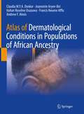
Atlas of Dermatological Conditions in Populations of African Ancestry
by Andrew F. Alexis Claudia M.Y.A. Donkor Jeannette Aryee-Boi Itohan Roseline Osazuwa Francis Kwame AffluThis book presents the nuances of dermatology from the African diaspora and the tropics. It not only addresses the dark pigmentation of the patient’s skin and the occurrence of tropical infections, but also the socioeconomic conditions which lead to unique features and the development of skin diseases. Chapters present numerous dermatological cases, with clear/relevant pictures, to serve as illustration of how skin conditions present in African/dark skin. Of these specific conditions, the book includes chapters on eczema, bullous diseases, hair disorders, acne, and papulosquamous disorders. Additionally, chapters address emotionally sensitive and socioeconomic-related issues such as skin bleaching and dermatological manifestations of HIV/AIDS, an infectious condition that disproportionately affects those residing in sub-Saharan Africa. Expertly written text is supplemented by hundreds of high-quality, real patient photos. Written by doctors living and treating patients in the tropical environment of Africa, Atlas of Dermatological Conditions in Populations of African Ancestry is an essential tool in broadening the scope of care for professionals and residents in dermatology alike.
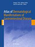
Atlas of Dermatological Manifestations of Gastrointestinal Disease
by George Y. Wu, Nathan Selsky and Jane M. Grant-KelsAtlas of Dermatological Manifestations of Gastrointestinal Disease provides a comprehensive compendium of digestive tract diseases with dermatological manifestations. The work is arranged by digestive symptoms, with sections based on the specific digestive disease etiology. Each section contains paired chapters of text, one on gastrointestinal manifestations and the other on dermatological manifestations. The atlas includes clinical presentations, pathophysiology, differential diagnosis, diagnostic tests/procedures, pathology, and references presented with substantial numbers of images. Concise and easy to use, Atlas of Dermatological Manifestations of Gastrointestinal Disease is an important resource for gastroenterologists, dermatologists, and internists alike in the treatment of digestive tract diseases.
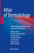
Atlas of Dermatology: Inflammatory, Infectious and Tumoral Skin Diseases
by Adriana Motta Luis Fernando González Gonzalo García Jennifer Guzmán Lorena Prada Hugo Herrera Mariam RolonDermatology is the science responsible for the study of the skin, mucous membranes (oral and genital) and cutaneous appendages, while dermatopathology focuses on its microscopic study. Although the two fields are closely related, in many cases the identification of dermatological diseases is mainly clinical and depends on the physician’s ability and experience. The purpose of this atlas, which collects over 900 clinical and histological photographs in high resolution, is to illustrate and describe the most frequent skin diseases on the basis of clinical cases. Offering a complete guide to the etiology, epidemiology, clinical features, histologic findings and diagnosis of the main skin diseases divided into three subgroups (inflammatory, infectious, or tumoral), it represents an invaluable resource for all medical students, residents, clinicians, and investigators learning dermatology.
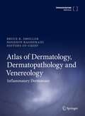
Atlas of Dermatology, Dermatopathology and Venereology: Cutaneous Anatomy, Biology And Inherited Disorders, And General Dermatologic Concepts
by Bruce Smoller Nooshin Bagherani
Atlas of Dermatology in Internal Medicine
by Néstor P. P. SánchezAtlas of Dermatology in Internal Medicine is the only concise text-atlas to cover the most common and most important cutaneous manifestations of systemic disease in children and adults. It features more than 150 clinical photographs that are accompanied by format-driven, clinically focused text on the diagnosis and management of cutaneous manifestations of connective tissue, pulmonary, renal, GI, endocrine, malignant, infectious, and HIV disease. There is also a separate chapter on skin diseases commonly seen in the ICU. A special feature is its systematic coverage of clinically relevant dermatopathology. The book is a helpful tool for physicians and trainees in internal medicine, family medicine, pediatrics, emergency medicine, and critical care medicine, as well as family, emergency, and critical care nurse practitioners.
Atlas of Dermatopathology: Practical Differential Diagnosis by Clinicopathologic Pattern
by Gunter Burg Werner Kempf Heinz KutznerImprove your dermatological diagnostic accuracy. The most efficient method in the diagnosis of skin diseases is the combined validation of clinical and histopathological features. The results of biopsy investigation in isolation don’t always yield answers. But when considered together, the clinical pattern and the histopathology, then diagnosis can become clearer. In this process the diagnostic impact of histopathology may be decisive or just confirmative to the clinical differential diagnosis. The structure of the book follows a basic approach to morphology, which is overall orientation at scanning magnification first, then identifying a prototypic pattern, and finally finding the essential diagnostic clue(s) under high power magnification. Dermatopathology: Practical Differential Diagnosis by Clinicopathologic Pattern provides top quality images to correlate clinical presentations with histopathologic features. Annotated images highlight subtle indications that can clinch the diagnosis. Concise, bullet-pointed text provides further context. Written by internationally renowned authors, the book is ideal for anyone involved in the diagnosis of skin disease.
Atlas of Dermatopathology: Practical Differential Diagnosis by Clinicopathologic Pattern
by Günter Burg Werner Kempf Heinz Kutzner Josef Feit Laszlo KaraiImprove your dermatological diagnostic accuracy. The most efficient method in the diagnosis of skin diseases is the combined validation of clinical and histopathological features. The results of biopsy investigation in isolation don’t always yield answers. But when considered together, the clinical pattern and the histopathology, then diagnosis can become clearer. In this process the diagnostic impact of histopathology may be decisive or just confirmative to the clinical differential diagnosis. The structure of the book follows a basic approach to morphology, which is overall orientation at scanning magnification first, then identifying a prototypic pattern, and finally finding the essential diagnostic clue(s) under high power magnification. Dermatopathology: Practical Differential Diagnosis by Clinicopathologic Pattern provides top quality images to correlate clinical presentations with histopathologic features. Annotated images highlight subtle indications that can clinch the diagnosis. Concise, bullet-pointed text provides further context. Written by internationally renowned authors, the book is ideal for anyone involved in the diagnosis of skin disease.
Atlas of Dermatopathology: Tumors, Nevi, and Cysts
by Gunter Burg Heinz Kutzner Werner Kempf Josef Feit Bruce R. SmollerDifferential diagnosis is at its most accurate and efficient when clinical presentation and histopathological features are considered in correlation with one another. With this being so, the expert team behind this atlas has integrated both perspectives to create an innovative and essential resource for all those involved with the diagnosis of tumors, cysts, and nevi. Almost 1,400 full-color images clearly illustrate common patterns and variants of tumorous lesions of the skin and are helpfully contextualized by concise, straightforward descriptions of key features and diagnostic clues.Whether they are aspiring or experienced practitioners, dermatologists and pathologists of all levels will find this an insightful and practically applicable addition to their bookshelf. Its far-reaching and easy-to-navigate coverage of relevant diseases of the skin provides trainees with an excellent grounding in the area, while practicing specialists may benefit from its use as a tool for the differential diagnoses of borderline cases.Atlas of Dermatopathology: Tumors, Nevi, and Cysts offers a uniquely balanced, clear, and comprehensive guide to what can be a difficult process, and will be of tremendous assistance tostudents, dermatologists, dermatopathologists, and pathologists everywhere. Atlas of Dermatopathology: Tumors, Nevi, and Cysts offers a uniquely balanced, clear, and comprehensive guide to what can be a difficult process, and will be of tremendous
Atlas of Dermatoscopy Cases: Challenging and Complex Clinical Scenarios
by Horacio Cabo Aimilios LallasThis practical atlas describes the use of dermatoscopy in the clinic, a technique that is increasingly used by the clinical dermatologist. It revolves around the use of clinical cases, simulating what happens in the clinic when the dermatologist is presented with a patient who has pigmented and non-pigmented lesions. Dermatologists perform diagnoses based on what they see on the skin and with these images recognize different diseases. This whole spectrum of forms and shapes is reflected in color. Dermatoscopy opens a new and very wide field of structures and colors that cannot be seen with the naked eye and, with appropriate training and the use of this book, improves clinical diagnosis. Atlas of Dermatoscopy Cases: Challenging and Complex Clinical Scenarios adds significantly to the analysis of cases from the Editors’ previous volume Comprehensive Atlas of Dermatoscopy Cases by teaching the technique through specially selected, complex, and interesting clinical cases and providing the reader a thorough understanding of the techniques and methodologies associated with diagnosis using dermatoscopy. It is of great use to the trainee dermatologist and any practicing dermatologist seeking to expand their skills with this important diagnostic tool.
Atlas of Dermatoses in Pigmented Skin
by Ranthilaka R. Ranawaka Ajith P. Kannangara Ajith KarawitaThis book focuses on the representation of dermatological diseases in pigmented skin, fills an important gap in the literature, and facilitates better dermatological diagnosis when dealing with patients of various ethnicities. It discusses over 400 diseases and their representations with the help of over 2000 high-quality images and illustrations. This book elaborates on each single disease using easy-to-follow schematics and a focused approach to facilitate reader learning. Pursuing a comprehensive, the book covers not only common skin diseases such as psoriasis, lichen planus, eczema, erythrasma, cutaneous tuberculosis, leprosy, leishmaniasis and oral submucous fibrosis, but also rare tropical diseases such as sporotrichosis, mycetoma, rhinosporodiosis, lobomycosis, mucormycosis and subcutaneous zygomycosis. It also addresses aesthetic concerns by covering hypopigmented and hyperpigmented disorders in pigmented skin such as guttate hypomelanosis, vitiligo, progressive macular hypomelanosis, chemical or physical induced depigmentation, melasma, sebo-melanosis, lichen and macular amyloidosis. Offering comprehensive coverage of dermatological disorders and diseases in pigmented skin, the book is a must-have resource for dermatology trainees and practitioners who treat or care for pigmented skin patients.
Atlas of Dermoscopy: Third Edition
by Ashfaq A Marghoob Ralph Braun Natalia JaimesThe much awaited third edition of the leading reference book in dermoscopy has undergone comprehensive revisions to all chapters, with updates and expanded content providing the reader with a more comprehensive and in-depth coverage of skin conditions, ranging from skin neoplasia to hair, nails, infections and inflammatory diseases. This compilation of contemporary dermoscopy knowledge will benefit the novice in building expertise and benefit experienced practitioners also by providing nuanced insights. Prepare to embark on a journey of learning as leading international experts in the field of dermoscopy explain via text and annotated exemplar images the amazing world of subsurface cutaneous colors and structures visible through a dermatoscope!
Atlas of Dermoscopy: Third Edition
by Ashfaq A. Marghoob Ralph Braun Natalia JaimesThe much awaited third edition of the leading reference book in dermoscopy has undergone comprehensive revisions to all chapters, with updates and expanded content providing the reader with a more comprehensive and in-depth coverage of skin conditions, ranging from skin neoplasia to hair, nails, infections and inflammatory diseases. This compilation of contemporary dermoscopy knowledge will benefit the novice in building expertise and benefit experienced practitioners also by providing nuanced insights. Prepare to embark on a journey of learning as leading international experts in the field of dermoscopy explain via text and annotated exemplar images the amazing world of subsurface cutaneous colors and structures visible through a dermatoscope!