- Table View
- List View

Color Atlas of Farm Animal Dermatology
by Danny W. ScottA practical full-color reference on the recognition and diagnosis of dermatological disease in farm animals Color Atlas of Farm Animal Dermatology, Second Edition is an essential reference for veterinary dermatologists, practitioners, and students alike. Emphasizing recognition and diagnosis, the book combines hundreds of color photographs with clear and concise text highlighting clinical features, differentials, and diagnostic information for each disease. Coverage includes both common and uncommon diseases of cattle, goats, sheep, llamas and alpacas, and pigs. This new edition has been fully updated throughout, and now includes information on llamas and alpacas. Diseases covered include bacterial, fungal, parasitic, viral and protozoal, immunological, congenital and hereditary, environmental, nutritional and miscellaneous diseases, as well as neoplastic and non-neoplastic growths. Offers a practical quick reference to aid in the diagnosis of dermatological conditions in farm animals, including cattle, goats, sheep, pigs, alpacas, and llamas Contains more than 700 full-color images Covers all dermatological disease types, organized by species-specific sections Thoroughly updated and now including coverage of camelids Color Atlas of Farm Animal Dermatology, Second Edition is a must-have book for veterinary dermatologists, practitioners, and students who work on farm animals.

Color Atlas of Human Poisoning and Envenoming
by Diaz JamesThe Color Atlas of Human Poisoning and Envenoming is the only full-color resource available for the immediate visual association and the rapid identification of envenoming species, resultant lesions, clinical outcomes of envenoming or poisoning, and recommended treatment strategies to limit toxic exposures and injuries. With no-nonsense, bulleted t
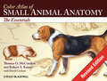
Color Atlas of Small Animal Anatomy: The Essentials
by Thomas O. McCracken Robert A. KainerThis new resource provides a basic foundation in small animal anatomy for students of veterinary medicine, animal science, and veterinary technology. Extraordinary accuracy and beautiful original artwork make this a truly unique learning tool that includes the anatomy of all organ systems in the dog, cat, rabbit, rat, and guinea pig - all described in a consistent manner. Learning features include: carefully selected labeling helps students learn and remember structures and relationships; male and female of species are depicted on facing pages so topographic anatomy can be compared; structures common to various animals are labeled several times, whereas unique structures are labeled on one or two species so students can make rapid distinctions of the structures peculiar to certain animals; and an introduction that provides readers with a background in nomenclature and anatomic orientation so they can benefit from the atlas even if they lack training in anatomy. The Atlas depicts topographic relationships of major organs in a simple, yet technically accurate presentation that's free from extraneous material so that those using the atlas can concentrate on the essential aspects of anatomy. It will be an invaluable resource for veterinary students, teachers and practitioners alike.
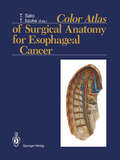
Color Atlas of Surgical Anatomy for Esophageal Cancer
by Tatsuo Sato and Toshifumi IizukaIt is essential to know all of the intricate lymph pathways when performing surgery for esophageal cancer in order to determine the extent of lymph node metastasis. Professor Sato has undertaken, at the request of the TNM Research Committee of the International Society for Diseases of the Esophagus, to map out and classify the lymph nodes of the mediastinum and neck. The beautiful artwork in the Color Atlas of Surgical Anatomy for Esophageal Cancer edited by Professor Sato gives an excellent understanding of the lymph node pathways and their importance in surgical treatment. Minute dissections which represent real life situations, not just the superficial pathways, show the precise location and topographical arrangement of the lymphatics. Full-color schematics are given with the actual dissection illustrations and photographs. The atlas clearly presents the classification of four significant pathways and their communication, the relationship of these pathways en route to the venous angles and the definition and assessment of the most critical nodes. Thoracic surgeons especially will benefit from the excellent illustrations of surgical techniques and the methods for recording the dissected lymph nodes which are presented by Professor Kakegawa. Leading experts fighting esophageal cancer with surgical treatment can use the classification in this outstanding atlas for many years to come as a standard for international comparison. The careful dissection of the lymph nodes may be the best way to improve survival rates after surgery for cancer of the thoracic esophagus.
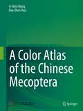
A Color Atlas of the Chinese Mecoptera
by Ji-Shen Wang Bao-Zhen HuaIn this atlas, 242 species of the Chinese Mecoptera are illustrated. For most species, the present pictures of the adults and the male genitalia are provided and a geographical distribution map is also available for every species. This book is aiming at not only the audiences such as general entomological researchers and hobbyists but also the forefront workers in the disciplines of agriculture and forestry as a practical reference.
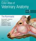
Color Atlas of Veterinary Anatomy, Volume 1, The Ruminants E-Book
by Raymond R. Ashdown Stanley H. Done Stephen W. BarnettThe Color Atlas of Veterinary Anatomy volume 1 presents a unique photographic record of dissections showing the topographical anatomy of the ruminant. With this book you will be able to see the position and relationships of the bones, muscles, nerves, blood vessels and viscera that go to make up each region of the body and each organ system. Each book in this three volume series is packed with full-color photographs and drawings of dissections prepared specifically for these texts.Accessibly and systematically structured with each chapter devoted to a specific body region.Important features of regional and topographical anatomy presented using full-color photos of detailed dissections.Detailed color line drawings clarify the relationships of relevant structures.Presents anatomy in a clinical context.Accompanying website with interactive quizzes and the chance to test yourself with self-assessment questions.New chapter on radiological anatomy.Special notes highlight clinical significance of each section.

Color Atlas of Veterinary Anatomy, Volume 2, The Horse - E-BOOK
by Raymond R. Ashdown Stanley H. DoneThe Color Atlas of Veterinary Anatomy volume 2 presents a unique photographic record of dissections showing the topographical anatomy of the horse. With this book you will be able to see the position and relationships of the bones, muscles, nerves, blood vessels and viscera that go to make up each region of the body and each organ system. Each book in this 3 volume series is packed with full-color photographs and drawings of dissections prepared specifically for these texts.Key features Accessibly and systematically structured with each chapter devoted to a specific body region Important features of regional and topographical anatomy presented using full color photos of detailed dissections Dissections presented in the standing position Detailed color line drawings clarify the relationships of relevant structures Presents anatomy in a clinical context This new edition second edition offers important new features, including: Accompanying website presents over 100 interactive quizzes and self-assessment questions Many more radiographs throughout Additional CT and MRI images Clinical notes highlight areas of particular clinical significance

Color Atlas of Veterinary Anatomy, Volume 3, The Dog and Cat E-Book (Color Atlas Of Veterinary Anatomy Ser. #Vol. 3)
by Stanley H. Done Peter C. Goody Susan A. Evans Neil C. SticklandIf you are looking for a book that presents a unique photographic record of dissections showing the topographical anatomy of the dog and cat: this is the atlas for you! Part of a comprehensive 3-volume set that also covers Ruminants (Volume 1) and The Horse (Volume 2), the Color Atlas of the Dog and Cat takes a complete look at virtually every aspect of veterinary anatomy. With this book you will be able to see the position and relationships of bones, muscles, nerves, blood vessels and viscera that go to make up each region of the body and each organ system. Rich with full-color photographs and drawings of dissections prepared specifically for these texts, each book in the series illustrates regional surface features photographed before dissection, then gives high-quality complementary photographs of articulated skeletons.Accessibly and systematically structured with each chapter is devoted to a specific body regionImportant features of regional and topographical anatomy presented in full color photos of detailed dissectionsDetailed color line drawings clarify the relationships of relevant structuresWebsite offers drag and drop quizzes and the chance to test yourself with mcqsInformative captions give additional information necessary for proper interpretation of the imagesPresents anatomy in a clinical context
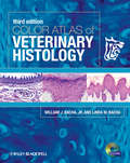
Color Atlas of Veterinary Histology
by Linda M. Bacha William J. Bacha Jr.Designed to provide students with a foundation in understanding and interpreting histologic and cytologic preparations, Color Atlas of Veterinary Histology is a practical benchside reference focusing on the normal histology of eight common domestic species. This Third Edition has been revised with new images, information, and updated terminology throughout. Introductory chapters have also been expanded to offer more complete coverage of the basic types of tissues, providing an even more thorough grounding in the principles of histology. For the first time, the more than 900 photomicrographs are available digitally in an interactive atlas on CD, offering images available for download with zoom capability. The new edition of this veterinary-specific histology atlas provides veterinary and veterinary technician students with an essential pictorial resource for interpreting histologic preparations.
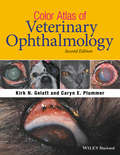
Color Atlas of Veterinary Ophthalmology
by Kirk N. Gelatt Caryn E. PlummerColor Atlas of Veterinary Ophthalmology, Second Edition provides a compendium of the clinical appearance of ophthalmic diseases likely to be encountered in small, large, or exotic animal practice. Offers a pictorial reference to the clinical appearance of diseases and conditions of the animal eye Presents multiple presentations of most ophthalmic diseases to show the varying ways the condition might appear Provides more than 1,000 high-quality color clinical photographs showing ocular disorders Includes new introductory chapters on ocular anatomy, the ophthalmic exam, and clinical findings in place of the clinical signs chapter Covers clinical history, the clinical signs and findings associated with the disease, the rule-outs or differential diagnoses, the recommended treatment, and the prognosis for each disorder
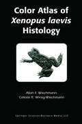
Color Atlas of Xenopus laevis Histology
by Allan F. Wiechmann Celeste R. Wirsig-WiechmannThe Color Atlas of "Xenopus laevis" Histology provides the first central source on the microscopic anatomy of cells, tissues, and major organs of the adult South African clawed frog, Xenopus laevis. For many years, X. laevis has been a highly popular experimental animal model in many areas of research. The recent development of transgenic Xenopus technology offers the promise that this animal model will be utilized more than ever before. The purpose of this book is to provide the active researcher with a central source of high quality light microscopic color images of the tissues of X. laevis, to aid in the identification of the cells and tissues of interest.
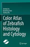
Color Atlas of Zebrafish Histology and Cytology
by Jianhua Hu Jinxing Lin Qiusheng ChenThis book elucidates the tissue structure and cell composition of the organs of zebrafish at the microscopic, ultrastructural and molecular levels. The distribution of important macromolecular substances is shown and the morphological relationship between different components is analyzed. The book is divided into 15 chapters and contains more than 700 structural photos, all of which are original experimental pictures of the research group. It shows the histological panorama of the whole zebrafish both in cross and longitudinal sections and covers and interprets the tissues and organs of zebrafish in detail, including oropharynx, taste buds, pharyngeal teeth, liver, etc. A brief text description of the structure and function meaning is available for every picture to facilitate the audience understanding the theoretical knowledge more vivid and concrete. In addition, the 3D reconstruction of the main organs of zebrafish is completed by computer-aided technology, and the three-dimensional morphology of the organs is displayed in an intuitive form. This book provides a reference for postgraduates and researchers in anatomy, biology, animal medicine, animal science, aquaculture, developmental biology, medicine, and experimental animals.
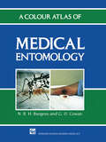
A Colour Atlas of Medical Entomology
by Nicholas Burgess G.O. CowanA there it is! guide to insects of medical and public health concern, mainly in the tropics. Each chapter covers identification, life cycle and habits of the causative stage and its medical/public health significance.
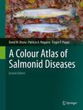
A Colour Atlas of Salmonid Diseases
by David W. Bruno Patricia A. Noguera Trygve T. PoppeSalmonids have widespread economic and environmental importance. Correct identification and understanding of their diseases are therefore vital if valuable stocks are to be maintained. This volume provides a practical guide and an aid to disease recognition. This is an updated and extended version of the first publication in 1996 and contains around 400 high quality colour photomicrographs.
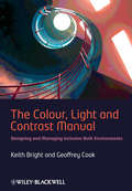
The Colour, Light and Contrast Manual: Designing and Managing Inclusive Built Environments
by Keith Bright Geoffrey CookEndorsed by the Society of Light and Lighting, this practical book offers comprehensive guidance on how colour, light and contrast can be incorporated within buildings to enhance their usability. The book provides state-of-the-art, clear guidance as well as a valuable information source for busy professionals involved in the design or management of new and existing environments. The ways colour, light and contrast are used within built environments are critical in determining how people interact with the space, and how confident, safe, and secure they will feel when doing so. They also have a major influence on a person’s sense of well-being and their ability to use the environment independently and without undue effort. Understanding how to use colour and contrast and how they are influenced by both natural and artificial lighting is vital for all those involved in the design and management of the environments and spaces we all use. In recent years there has been a considerable amount of work undertaken to further our understanding of how colour, light and contrast affect emotion and sensory abilities, and how they can assist or hinder people in their everyday lives. Other publications consider these issues individually but The Colour, Light and Contrast Manual: designing and managing inclusive built environments draws knowledge and information together to produce a unique, comprehensive and informative guide to how the three elements can work together to improve the design and management of environments for us all. Supporting website at: www.wiley.com/go/brightandcook
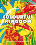
Colourful Kingdom: How animals use colour to surprise and survive
by Anna OmedesGet familiar with animals from around the world who use colour as a super power! In this fact-filled and engaging book, you will read surprising stories of how animals use colour to thrive in their habitat.Colour gives them unique powers: the giant cuttlefish changes colour to hide from predators, whilst the firefly glows in the dark to choose a mate; and if the flamingo was to stop eating brine shrimp, they would no longer be pink!Learn about the venomous spiders from the jungle, how tigers use camouflage to hunt their preys or how coral and fish have learned to live in harmony with each other.Each page is vividly illustrated and is accompanied by authoritative text, providing an exploration of the animal's habitats and habits.
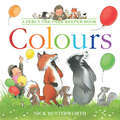
Colours (Percy the Park Keeper)
by Nick ButterworthLearn the colours of the rainbow with Percy the park keeper and his animal friends in this fun first concept book from highly regarded, award-winning author/illustrator Nick Butterworth, creator of One Snowy Night.
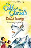
Colt of the Clouds (The Winged Horse Race #2)
by Kallie GeorgeJourney to the clouds and back in Colt of the Clouds, the breathtaking second Winged Horse Race adventure from Kallie George, illustrated by Lucy Eldridge.Ever since Pippa and her beloved horse, Zephyr, were banished from the slopes of Mount Olympus, they’ve tried to adjust to living a normal life on the ground. But when Pippa rescues a winged colt, she knows she must take return him to the land of the gods and goddesses. Up on Olympus, however, disaster awaits. The gods and goddesses are at war, and all of the winged horses have disappeared.With the help of a new friend, Hero, Pippa takes on a task few mortals would dare: to find the winged horses and end the war. Join the winged horses in this fast-paced story of loyalty, friendship and bravery.
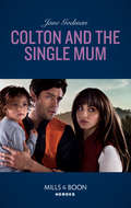
Colton And The Single Mum (The Coltons of Red Ridge #4)
by Jane GodmanA ready made family…A serial killer on the loose
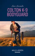
Colton K-9 Bodyguard: Colton K-9 Bodyguard Conard County Revenge Killer Secrets Second Chance Soldier (The Coltons of Red Ridge #3)
by Lara LacombeHe broke her heart, now he’s back to save her.
Colton K-9 Target: Dead In The Water / Colton K-9 Target (the Coltons Of Grave Gulch) (The Coltons of Grave Gulch #8)
by Justine DavisMan's best friend and most trusted partner
Colton P.i. Protector: Colton P. I. Protector The Texas Soldier's Son The Fugitive's Secret Child Snowbound Security (The Coltons of Red Ridge #5)
by Regan BlackHe’s claiming redemption And the woman he wants!